Bacillus thuringiensis toxin in C. elegans: Difference between revisions
Adrianowyczs (talk | contribs) |
Adrianowyczs (talk | contribs) |
||
| Line 3: | Line 3: | ||
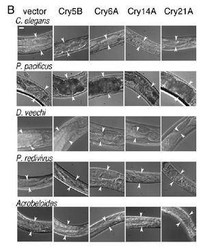
of the anterior intestine of nematodes fed the four toxic crystal proteins in E. coli. Arrowheads delineate the width of the intestine at one position near the | of the anterior intestine of nematodes fed the four toxic crystal proteins in E. coli. Arrowheads delineate the width of the intestine at one position near the | ||
anterior. (Bar 5 20 mm.)]] | anterior. (Bar 5 20 mm.)]] | ||
[[Image:Wei2003_1.jpeg|thumb|300px|right| | [[Image:Wei2003_1.jpeg|thumb|300px|right|Figure 1. Infection of C. elegans by B. thuringiensis and B. anthracis. Top row: Dissecting microscope view of nematodes cultured under various | ||
of the anterior | conditions. Scale bar of all images in top row is 500 mm. Bottom row: Compound microscope view of nematodes cultured under various conditions. | ||
For all images in the bottom row, anterior of the worm is top right and scale bar is 50 mm. (A) C. elegans cultured in a well with B. thuringiensis without | |||
Cry5B. Top row: None of the six nematodes are infected. All are healthy. The blur associated with some of the worms in the top row is due to their | |||
movement in the well. Bottom row: The internal structures of C. elegans fed B. thuringiensis without Cry5B, including the pharynx and intestine, are all | |||
intact. (B) C. elegans cultured in a well with B. thuringiensis and Cry5B. Top row: Five of the six worms are completely infected (rigid, lack of internal | |||
structures and normal coloration); one is not. Bottom row: Infected animals show complete or near complete digestion of internal structures by the | |||
bacteria. Vegetative and sporulated bacteria can be seen in these lethally infected animals. (C) Similar images as in (B) except the bacterium cultured | |||
with the nematodes is Bacillus anthracis.]] | |||
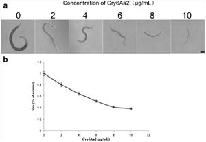
[[Image:Luo2013_1.jpg|thumb|300px|right|Growth assay of L1 larvae | [[Image:Luo2013_1.jpg|thumb|300px|right|Growth assay of L1 larvae | ||
of C. elegans with Cry6Aa2 | of C. elegans with Cry6Aa2 | ||
Revision as of 00:01, 22 April 2014
Introduction


By Sarah Adrianowycz
At right is a sample image insertion. It works for any image uploaded anywhere to MicrobeWiki. The insertion code consists of:
Double brackets: [[
Filename: PHIL_1181_lores.jpg
Thumbnail status: |thumb|
Pixel size: |300px|
Placement on page: |right|
Legend/credit: (B) Photographs
of the anterior intestine of nematodes fed the four toxic crystal proteins in E. coli. Arrowheads delineate the width of the intestine at one position near the
anterior. (Bar 5 20 mm.)
Closed double brackets: ]]
Other examples:
Bold
Italic
Subscript: H2O
Superscript: Fe3+
Introduce the topic of your paper. What microorganisms are of interest? Habitat? Applications for medicine and/or environment?
Section 1
Include some current research, with at least one figure showing data.
