Utilization of Bacillus thuringiensis in Genetically Modified Crops: Difference between revisions
No edit summary |
No edit summary |
||
| Line 36: | Line 36: | ||
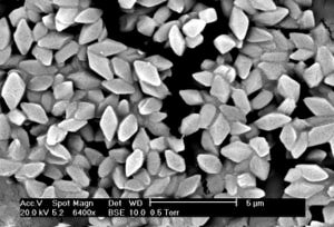
[[Image:Bacillus thuringiensis.JPG|thumb|300px|left|Figure 3. Electron micrograph of the crystalline protein toxin produced by <i>Bacillus thuringiensis</i>. The insecticidal properties of these proteins were first discovered by Christopher Hannay in 1955. Micrography by Jim Buckman (2006).]] | [[Image:Bacillus thuringiensis.JPG|thumb|300px|left|Figure 3. Electron micrograph of the crystalline protein toxin produced by <i>Bacillus thuringiensis</i>. The insecticidal properties of these proteins were first discovered by Christopher Hannay in 1955. Micrography by Jim Buckman (2006).]] | ||
Figure 3. <br> | Figure 3. <br> | ||
<br> | <br> | ||
| Line 45: | Line 41: | ||
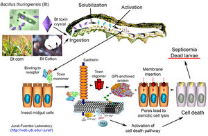
[[Image:Bt whole process.jpeg|thumb|300px|right|Figure 4.]] | [[Image:Bt whole process.jpeg|thumb|300px|right|Figure 4.]] | ||
Figure 4. <br> | Figure 4. <br> | ||
<br> | |||
==Ethical Issues Surrounding Bt Crops== | |||
Include some current research, with at least one figure showing data.<br> | |||
<br> | <br> | ||
Revision as of 20:32, 20 April 2015
Introduction
By Zoë Frazier
At right is a sample image insertion. It works for any image uploaded anywhere to MicrobeWiki. The insertion code consists of:
Double brackets: [[
Filename: PHIL_1181_lores.jpg
Thumbnail status: |thumb|
Pixel size: |300px|
Placement on page: |right|
Legend/credit: Electron micrograph of the Ebola Zaire virus. This was the first photo ever taken of the virus, on 10/13/1976. By Dr. F.A. Murphy, now at U.C. Davis, then at the CDC.
Closed double brackets: ]]
Other examples:
Bold
Italic
Subscript: H2O
Superscript: Fe3+
Introduce the topic of your paper. What microorganisms are of interest? Habitat? Applications for medicine and/or environment?
Structure and Phylogeny
Include some current research, with at least one figure showing data.
History
Include some current research, with at least one figure showing data.
Life Cycle
Figure 2.
Bt Toxins
Figure 3.
Bt Crops
Figure 4.
Ethical Issues Surrounding Bt Crops
Include some current research, with at least one figure showing data.
Evolved Resistance and Secondary Pests

Figure 5.
Conclusion
Include conclusion
References
[1] Ibrahim, M.A., N. Griko, M. Junker, and L.A. Bulla. 2010. Bacillus thuringiensis: A genomic and proteomics perspective. Bioengineered Bugs 1:1, 31-50.
[2]


