Campylobacter fetus: Difference between revisions
| Line 26: | Line 26: | ||
==Genome structure== | ==Genome structure== | ||
''Campylobacter Fetus'' was completely | ''Campylobacter Fetus'' was completely sequenced and was found to have 1820 genes. It has a circular DNA. Its genome length is 1,773,615 nt. GC content is 33%, coding sequence is 90%.It as 3441 proteins(7).The two stains that were found were dividing based on their lipopolysaccharide (LPS) composition. These unique stains must contain conserved sequences so to check their unique versus mutual sequences we used gel electrophoresis. This helped answer the unique method of this pathogen in changing its S layer proteins. | ||
The two stains that were found were dividing based on their lipopolysaccharide (LPS) composition. These unique stains must contain conserved sequences so to check their unique versus mutual sequences we used gel electrophoresis. This helped answer the unique method of this pathogen in changing its S layer proteins. | Different study (5) reported the different multiple highly conserved S layer homologous proteins, that are tightly packed in this species. These special S layer proteins are of the sizes of 97-149 kDa. They attach to the N terminus of the cell wall lipopolysaccharide non covalently. The Genome holds one promoter for these proteins; it has at least one open frame and a DNA inversion. The inversion is using homologous recombination. | ||
Different study reported the different multiple | |||
==Cell structure and metabolism== | ==Cell structure and metabolism== | ||
Revision as of 03:12, 27 May 2007
A Microbial Biorealm page on the genus Campylobacter fetus
Classification
Higher order taxa
Bacteria; Proteobacteria; Epsilon Proteobacteria; Campylobacterales; Campylobacteraceae; Campylobacter
Species
C. fetus
|
NCBI: Taxonomy |
Description and significance
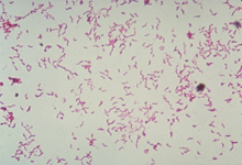
Campylobacter fetus is a spiral slender, spirally curved bacterial pathogen, enclosed with an S layer of special crystalline surface proteins. (Their unique function will be discussed later) . It is a gram negative species holding two membranes and a thin cell wall in between. Since it is a pathogen, it can reside anywhere in the human body. The campylobacter S layer proteins were found to have a virulence factor in resistance to the host immune defense mechanisms. Two subspecies were then suspected to exist in the campylobacter. One was Campylobacter fetus subsp. fetus and Campylobacter fetus subsp. Venerealis. To further investigate the genetic diversity among C. fetus strains of different origins, multiple genetic analyzing were used such as polymorphic DNA (RAPD), pulsed-field gel electrophoresis (PFGE), and DNA-DNA hybridization. I t was also mainly found that its natural habitat of C. fetus subsp. fetus is the intestinal tract of cattle, but it can also cause abortions.
Genome structure
Campylobacter Fetus was completely sequenced and was found to have 1820 genes. It has a circular DNA. Its genome length is 1,773,615 nt. GC content is 33%, coding sequence is 90%.It as 3441 proteins(7).The two stains that were found were dividing based on their lipopolysaccharide (LPS) composition. These unique stains must contain conserved sequences so to check their unique versus mutual sequences we used gel electrophoresis. This helped answer the unique method of this pathogen in changing its S layer proteins. Different study (5) reported the different multiple highly conserved S layer homologous proteins, that are tightly packed in this species. These special S layer proteins are of the sizes of 97-149 kDa. They attach to the N terminus of the cell wall lipopolysaccharide non covalently. The Genome holds one promoter for these proteins; it has at least one open frame and a DNA inversion. The inversion is using homologous recombination.
Cell structure and metabolism
It has flagella for motility using the proton motive force for energy. Since it is gram negative, it has three layer of protection – inner membrane, thin murien layer and an outer membrane. This organism has the optimal growth at atmospheres enriched with CO2 and 5% oxygen with nitrogen or hydrogen according to the journal of bacteriology(2). Camplylobacter fetus needs an anaerobic environment for growth. It needs a respiratory type of metabolism for growth. These bacteria do not ferment; they also do not oxidize carbohydrates. Their entire energy is produced by certain amino acids and the tricarboxlic acid cycle intermediates. Their special features are their unique S layer surface proteins.
Ecology
Campylobacter fetus has been studied and shown to adhere to those intestinal epithelial cell lines that it invades in humans. The information from this study reported in the Canadian Journal of microbiology (1) shows that this campylobacter fetus showed capabilities of adhering to many cell lines. This is just one example of the kind of cells this bacterium interacts with. Since this is a pathogen it interacts with many host cells either in humans or in animals mostly cattle.
Pathology
Campylobacter fetus is a bacterium that can interact and disrupt reproduction cycles. Since they have many possible surface layer proteins they have an antigenic variation that can trick the immune system. There are two types of mechanism in which these bacteria infect since there are two subspecies. The Campylobacter fetus subsp. Venerealis is found on bull mucus layer over the crypt cell layer of the male prepuce. This way, the bacteria is transmitted to the female cattle and causes its infertility. This is also the way it will get transferred again during mating. Campylobacter fetus subsp. Fetus is using a different infection mechanism in which the animal whether bird, reptile ingest the bacteria, invades the mucus cell and can cause an abortion. The two subspecies uses different ways to invade the host. Humans are mostly not affected but can accidentally ingest the Campylobacter fetus subsp. Fetus and suffer sever consciousness such as inflammation of intestinal tract.(5)
Application to Biotechnology
As mentioned before the unique S layer coat proteins provide the bacteria with resistant to detection by the host immune system since the proteins can change their shape and size. This is making this pathogen to be very dangerous to its host and vast research is made on characterizing these proteins. (8)
Current Research
- Extensive research is being done through the years as well as in current times dealing with the big mystery of the S layer proteins that escape the immune system. Dr. Thompson (9)
Suggest two mechanisms for this. One is the resistance to the non immune serum by preventing the binding of this serum to the C. fetus cell surface proteins. Another way might be the one I already mentioned about varying its S layer coat proteins thus avoid the antibodies of the immune system.
- Another recent research was made by Briedis (4) in which blood from a patient with cellulites showed evidence of C. fetus. Later on in testing, he found evidence for the other strain Venerealis. He continued with testing to show that the strains differ in their 1% glycine tolerance. More research is done by him to finalize the results and come with a more conclusive result as to what strain appeared in his patient.
- To find more about the two different strains of campylobacter fetus , Schulze (3) conducted some tests to examine the main differences between the two strains .He didn’t find more than what was already known of the two subspecies but run results that gave more light for the main differences using some tests as PCR, temperature sensitive , phenotype and genotype testing as well as many more.
References
1. Lori L Graham, “Campylobacter fetus adheres to and enters INT 407 cells” Canadian Journal of Microbiology 48 pp 995-1007
2. G M Carlone and J Lascelles, “Aerobic and anaerobic respiratory systems in Campylobacter fetus subsp. jejuni grown in atmospheres containing hydrogen.” Journal of Bacteriology, 1982 October; 152(1): 306–314.
3. Frank Schulze, Audrey Bagon, Wolfgang Müller, and Helmut Hotzel, “Identification of Campylobacter fetus Subspecies by Phenotypic Differentiation and PCR” Journal of Clinical Microbiology 2006 June; 44(6): 2019–2024.
4. Dalius J. Briedis, Ali Khamessan, Richard W. McLaughlin, Hojatollah Vali, Maria Panaritou, and Eddie C. S. Chan, “Isolation of Campylobacter fetus subsp. fetus from a Patient with Cellulitis” Journal of Clinical Microbiology, December 2002, p. 4792-4796, Vol. 40, No. 12
5. Joel Dworkin & Martin J. Blaser, “Molecular mechanisms of Campylobacter fetus surface layer protein expression.” Molecular Microbiology- Volume 26 Issue 03 Page 433 - October 1997
6. PAUL EDMONDS, CHARLOTTE M. PATTON, TIMOTHY J. BARRETT, GEORGE K. MORRIS, ARNOLD G.STEIGERWALT, AND DON J. BRENNER “Biochemical and Genetic Characteristics of Atypical Campylobacter fetus subsp. fetus Strains Isolated from Humans in the United States” JOURNAL OF CLINICAL MICROBIOLOGY, June 1985, p. 936-940 Vol. 21, No. 6
7. NCBI –National center for biotechnology information http://www.ncbi.nlm.nih.gov/entrez/query.fcgi?db=genome&cmd=Retrieve&dopt=Overview&list_uids=20090
8. E Wang, M M Garcia, M S Blake, Z Pei, and M J Blaser “Shift in S-layer protein expression responsible for antigenic variation in Campylobacter fetus.” Journal of bacteriology 1993 August; 175(16): 4979–4984.
9. Dr. Stuart A. Thompson “Campylobacter Surface-Layers (S-Layers) and Immune Evasion” Annals of Periodontology December 2002, Vol. 7, No. 1, Pages 43-53
Edited by Sharon Porath of Rachel Larsen and Kit Pogliano

