The Monoxenous Life Cycle Of Eimeria: Difference between revisions
No edit summary |
No edit summary |
||
| Line 4: | Line 4: | ||
<br>By Emma Stewart-Bates<br> | <br>By Emma Stewart-Bates<br> | ||
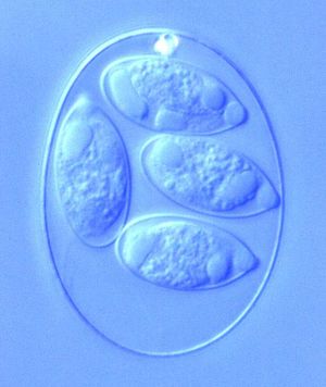
<br><i>Eimeria</i> is a genus of protozoa that are parasitic to many vertebrate animals, most often cattle, domesticated birds, goats, and sheep. These parasites contain an apical complexes and apicoplasts, organelles that allow the cell to enter a host organism. The life cycle of <i>Eimeria</i> is considered monoxenous, meaning that the cycle occurs in one host. The three stages of its life cycle include oocyst, sporozoite, and merozoite. They undergo both sexual and asexual reproduction during different stages of their life. Animals infected by <i>Eimeria</i> often develop the disease coccidiosis, which mainly causes diarrhea, fatigue, and loss of appetite. Coccidiosis is spread when an animal ingests infected tissue or is exposed to contaminated feces.<br> | <br><i>Eimeria</i> is a genus of protozoa that are parasitic to many vertebrate animals, most often cattle, domesticated birds, goats, and sheep. These parasites contain an apical complexes and apicoplasts, organelles that allow the cell to enter a host organism. The life cycle of <i>Eimeria</i> is considered monoxenous, meaning that the cycle occurs in one host. The three stages of its life cycle include oocyst, sporozoite, and merozoite. They undergo both sexual and asexual reproduction during different stages of their life. Animals infected by <i>Eimeria</i> often develop the disease coccidiosis, which mainly causes diarrhea, fatigue, and loss of appetite. Coccidiosis is spread when an animal ingests infected tissue or is exposed to contaminated feces.<ref>[http://parasite.org.au/para-site/text/eimeria-text.html "<i>Eimeria</i>." The Australian Society for Parasitology Inc., 16 June 2010. Web. 15 Apr. 2017.]</ref><br> | ||
<br>At right is a sample image insertion. It works for any image uploaded anywhere to MicrobeWiki.<br><br>The insertion code consists of: | <br>At right is a sample image insertion. It works for any image uploaded anywhere to MicrobeWiki.<br><br>The insertion code consists of: | ||
| Line 21: | Line 21: | ||
<br>Introduce the topic of your paper. What is your research question? What experiments have addressed your question? Applications for medicine and/or environment?<br> | <br>Introduce the topic of your paper. What is your research question? What experiments have addressed your question? Applications for medicine and/or environment?<br> | ||
Sample citations: | Sample citations: | ||
<ref>[http://www.ncbi.nlm.nih.gov/pmc/articles/PMC3847443/ Bartlett et al.: Oncolytic viruses as therapeutic cancer vaccines. Molecular Cancer 2013 12:103.]</ref> | <ref>[http://www.ncbi.nlm.nih.gov/pmc/articles/PMC3847443/ Bartlett et al.: Oncolytic viruses as therapeutic cancer vaccines. Molecular Cancer 2013 12:103.]</ref> | ||
<br><br>A citation code consists of a hyperlinked reference within "ref" begin and end codes. | <br><br>A citation code consists of a hyperlinked reference within "ref" begin and end codes. | ||
Revision as of 14:33, 15 April 2017
Introduction
By Emma Stewart-Bates
Eimeria is a genus of protozoa that are parasitic to many vertebrate animals, most often cattle, domesticated birds, goats, and sheep. These parasites contain an apical complexes and apicoplasts, organelles that allow the cell to enter a host organism. The life cycle of Eimeria is considered monoxenous, meaning that the cycle occurs in one host. The three stages of its life cycle include oocyst, sporozoite, and merozoite. They undergo both sexual and asexual reproduction during different stages of their life. Animals infected by Eimeria often develop the disease coccidiosis, which mainly causes diarrhea, fatigue, and loss of appetite. Coccidiosis is spread when an animal ingests infected tissue or is exposed to contaminated feces.[1]
At right is a sample image insertion. It works for any image uploaded anywhere to MicrobeWiki.
The insertion code consists of:
Double brackets: [[
Filename: PHIL_1181_lores.jpg
Thumbnail status: |thumb|
Pixel size: |300px|
Placement on page: |right|
Legend/credit: Electron micrograph of the Ebola Zaire virus. This was the first photo ever taken of the virus, on 10/13/1976. By Dr. F.A. Murphy, now at U.C. Davis, then at the CDC.
Closed double brackets: ]]
Other examples:
Bold
Italic
Subscript: H2O
Superscript: Fe3+
Introduce the topic of your paper. What is your research question? What experiments have addressed your question? Applications for medicine and/or environment?
Sample citations:
[2]
A citation code consists of a hyperlinked reference within "ref" begin and end codes.
Section 1
Include some current research, with at least one figure showing data.
Every point of information REQUIRES CITATION using the citation tool shown above.
Section 2
Include some current research, with at least one figure showing data.
Section 3
Include some current research, with at least one figure showing data.
Section 4
Conclusion
References
Authored for BIOL 238 Microbiology, taught by Joan Slonczewski, 2017, Kenyon College.

