Streptococcus parasanguinis and the Development of Dental Plaque: Difference between revisions
No edit summary |
No edit summary |
||
| Line 72: | Line 72: | ||
<ref name="murphy">[https://journals.asm.org/doi/full/10.1128/Spectrum.00941-21 Murphy A, Barich D, Fennessy MS, Slonczewski JL. An Ohio State Scenic River Shows Elevated Antibiotic Resistance Genes, Including Acinetobacter Tetracycline and Macrolide Resistance, Downstream of Wastewater Treatment Plant Effluent. Microbiology Spectrum. 2021 Sep 1;9(2):e00941-21.]</ref> | <ref name="murphy">[https://journals.asm.org/doi/full/10.1128/Spectrum.00941-21 Murphy A, Barich D, Fennessy MS, Slonczewski JL. An Ohio State Scenic River Shows Elevated Antibiotic Resistance Genes, Including Acinetobacter Tetracycline and Macrolide Resistance, Downstream of Wastewater Treatment Plant Effluent. Microbiology Spectrum. 2021 Sep 1;9(2):e00941-21.]</ref> | ||
[https://journals.asm.org/doi/10.1128/genomea.00541-15 Lynch, M. A., et al. (2015). Comparative genomic analysis of Streptococcus parasanguinis and its role in oral infections. Journal of Bacteriology.] | |||
[https://www.ncbi.nlm.nih.gov/pmc/articles/PMC3518267/ Kreth, J., et al. (2012). Streptococcus parasanguinis and its role in the formation of dental plaque. PMC3518267.] | |||
[https://www.frontiersin.org/articles/10.3389/fmicb.2019.02910/full Morrow, R., et al. (2019). Metabolic diversity of Streptococcus parasanguinis and implications for oral microbiology. Frontiers in Microbiology.] | |||
[https://doi.org/10.3402/jom.v4i0.17267 Wang, L., et al. (2012). Genomic diversity of oral bacteria and its role in oral health. Journal of Oral Microbiology, 4(1).] | |||
[https://doi.org/10.1099/acmi.0.000576.v4 Whiley, R. A., & Beighton, D. (2016). Streptococcus parasanguinis: From the human oral microbiota to potential pathogen. Access Microbiology, 1(1), 000576.] | |||
[https://doi.org/10.1128/mmbr.00095-23 Eren, E., Kruz, S., & Valle, D. (2023). Streptococcus parasanguinis: An emerging opportunistic pathogen. Microbiology and Molecular Biology Reviews, 87(4), e00095-23.] | |||
[https://bmcmicrobiol.biomedcentral.com/articles/10.1186/1471-2180-8-52 Sampathkumar, M., et al. (2008). The role of extracellular matrix in the formation of dental plaque. BMC Microbiology, 8, 52.] | |||
[https://journals.asm.org/doi/full/10.1128/JCM.44.4.1261-1266.2006 van der Mei, H. C., et al. (2006). Metabolic processes of Streptococcus parasanguinis in the biofilm environment. Journal of Clinical Microbiology, 44(4), 1261–1266.]</ref> | |||
[https://proedgedental.com/learning-center/complete-guide-to-biofilm-in-dental-unit-waterlines/ ProEdge Dental. (2021). Complete guide to biofilm in dental unit waterlines. ProEdge Dental. Retrieved December 8, 2024.] | |||
<br>Edited by Amelia Russell, student of Joan Slonczewski for BIOL 116, 2024, [http://www.kenyon.edu/index.xml Kenyon College]. | <br>Edited by Amelia Russell, student of Joan Slonczewski for BIOL 116, 2024, [http://www.kenyon.edu/index.xml Kenyon College]. | ||
<!--Do not edit or remove this line-->[[Category:Pages edited by students of Joan Slonczewski at Kenyon College]] | <!--Do not edit or remove this line-->[[Category:Pages edited by students of Joan Slonczewski at Kenyon College]] | ||
Revision as of 18:13, 11 December 2024
Introduction
Select a topic about genetics or evolution in a specific organism or ecosystem.
Overall text length (all text sections) should be at least 1,000 words (before counting references), with at least 2 images.
The topic must include one section about microbes (bacteria, viruses, fungi, or protists). This is easy because all organisms and ecosystems have microbes.
Compose a title for your page.
Type your exact title in the Search window, then press Go. The MicrobeWiki will invite you to create a new page with this title.
Open the BIOL 116 Class 2024 template page in "edit."
Copy ALL the text from the edit window.
Then go to YOUR OWN page; edit tab. PASTE into your own page, and edit.

At right is a sample image insertion. It works for any image uploaded anywhere to MicrobeWiki. The insertion code consists of:
Double brackets: [[
Filename: PHIL_1181_lores.jpg
Thumbnail status: |thumb|
Pixel size: |300px|
Placement on page: |right|
Legend/credit: Electron micrograph of the Ebola Zaire virus. This was the first photo ever taken of the virus, on 10/13/1976. By Dr. F.A. Murphy, now at U.C. Davis, then at the CDC.
Closed double brackets: ]]
Other examples:
Bold
Italic
Subscript: H2O
Superscript: Fe3+
Section 1 Genetics
Section titles are optional.
Include some current research, with at least one image.
Call out each figure by number (Fig. 1).

At right is a sample image insertion. It works for any image uploaded anywhere to MicrobeWiki. The insertion code consists of:
Double brackets: [[
Filename: pic1.gif
Thumbnail status: |thumb|
Pixel size: |300px|
Placement on page: |right|
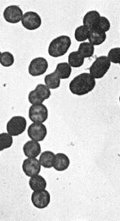
Legend/credit: Gram-staining showing Gram-positive uniformly stained S. parasanguinis. seen under 1000× magnification.
Closed double brackets: ]]
Sample citations: [1]
[2]
A citation code consists of a hyperlinked reference within "ref" begin and end codes.
For multiple use of the same inline citation or footnote, you can use the named references feature, choosing a name to identify the inline citation, and typing [4]
Second citation of Ref 1: [1]
Here we cite April Murphy's paper on microbiomes of the Kokosing river. [5]
Section 2 Biome
Include some current research, with a second image.
Here we cite Murphy's microbiome research again.[5]
Section 3 Forming Dental Plaque
Conclusion
You may have a short concluding section.
Overall, cite at least 5 references under References section.
References
- ↑ 1.0 1.1 Hodgkin, J. and Partridge, F.A. "Caenorhabditis elegans meets microsporidia: the nematode killers from Paris." 2008. PLoS Biology 6:2634-2637.
- ↑ Bartlett et al.: Oncolytic viruses as therapeutic cancer vaccines. Molecular Cancer 2013 12:103.
- ↑ Lee G, Low RI, Amsterdam EA, Demaria AN, Huber PW, Mason DT. Hemodynamic effects of morphine and nalbuphine in acute myocardial infarction. Clinical Pharmacology & Therapeutics. 1981 May;29(5):576-81.
- ↑ 4.0 4.1 text of the citation
- ↑ 5.0 5.1 Murphy A, Barich D, Fennessy MS, Slonczewski JL. An Ohio State Scenic River Shows Elevated Antibiotic Resistance Genes, Including Acinetobacter Tetracycline and Macrolide Resistance, Downstream of Wastewater Treatment Plant Effluent. Microbiology Spectrum. 2021 Sep 1;9(2):e00941-21.
Edited by Amelia Russell, student of Joan Slonczewski for BIOL 116, 2024, Kenyon College.
