Ophiocordyceps unilateralis: Difference between revisions
| Line 68: | Line 68: | ||
The evidence from fossil samples those taken from the Messel Pit in eastern Germany of leaves from Byttnertiopsis daphnogenes when compared to leaves in the Peninsula Khao Chong Botanic Garden in Thailand shows similar bite patterns or “death grip,” of ants infected with Ophiocordyceps unilateralis. The unique bite patterns of the leaves from the Messel Pit were originally believed to be typical vein-cutting behavior common to herbivorous insects. However this hypothesis was discounted when the bite patterns did not match the usual patterns associated with vein-cutting. Instead the bite marks distinctively resembled those of ants infected with O. unilateralis, with the addition that the marks were around major veins. Radiometric dating of the leaves show that they are around 47 million years old, which provides evidence that the parasite manipulation found in O. unilateralis is one of the oldest interactions documented [4]. | The evidence from fossil samples those taken from the Messel Pit in eastern Germany of leaves from Byttnertiopsis daphnogenes when compared to leaves in the Peninsula Khao Chong Botanic Garden in Thailand shows similar bite patterns or “death grip,” of ants infected with Ophiocordyceps unilateralis. The unique bite patterns of the leaves from the Messel Pit were originally believed to be typical vein-cutting behavior common to herbivorous insects. However this hypothesis was discounted when the bite patterns did not match the usual patterns associated with vein-cutting. Instead the bite marks distinctively resembled those of ants infected with O. unilateralis, with the addition that the marks were around major veins. Radiometric dating of the leaves show that they are around 47 million years old, which provides evidence that the parasite manipulation found in O. unilateralis is one of the oldest interactions documented [4]. | ||
<iframe width="560" height="315" src="http://www.youtube.com/embed/XuKjBIBBAL8" frameborder="0" allowfullscreen></iframe> | |||
==References== | ==References== | ||
Revision as of 04:47, 28 February 2012
A Microbial Biorealm page on the genus Ophiocordyceps unilateralis
Classification
| Fungi | Ascomycota | Sordariomycetes | Hypocreales | Ophiocordycipitaceae | Ophiocordyceps | O. unilateralis | Ophiocordyceps unilateralis
Description and Significance
Ophiocordyceps unilateralis (Ascomycota: Hypocreales) is a specialized fungal parasite that infects, manipulates and kills formicine ants, predominantly in tropical forest ecosystems. It specifically infects Camponotus leonardi of the tribe of campotini [3, 2].
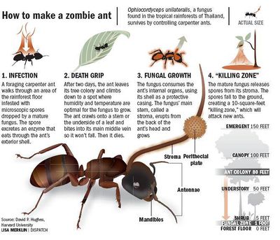
Worker ants are infected during foraging when the fungal spores attach to their cuticles [Hughes]. Germination and then penetration through the cuticle leads to rapid infection inside the host body [1, 2]. Once infected the ants will climb down from their natural habitats on rainforest tree and relocate to 25 cm off the ground under leaves where the temperature is low and humidity is high. Fungal reproduction is only possible after a stalk is grown out of the host’s head by propulsion of spores out from its fruiting bodies [3]. Spores of O. unilateralis are actively discharged and dispersed over short distances, creating an infectious “killing field” of ∼1 m2 below the dead host (N. L. Hywel-Jones, unpublished data)[10].
Fungal manipulation of an ant host’s mouthparts was found on a 48 million year old single leaf of the dicotyledonous plant host Byttnertiopsis daphnogenes from Messel in Northern Germany [4]. The close modern parallel for this distinctive type of leaf damage is the death grip of some fungus-infected carpenter ants such as the fungus O. unilateralis which adaptively manipulates worker ants of C. leonardi to bite along major veins of leaves in Thai tropical forests. This is the oldest evidence of parasites manipulating the behaviour of their hosts and suggests that the specialized interaction is relatively ancient rather than newly acquired[5].
Due to the increased amount of research on Ophiocordyceps in recent years, the name Ophiocordyceps unilateralis is often extended to Ophiocordyceps unilateralis sensu lato to indicate that the taxonomic system for this species is currently in flux [2] and will change as the genus and species characteristics become more defined.
Genome Structure
The genome of this organism is not sequenced entirely, although major enzymes such as RNA polymerases have been sequenced. Four polyketide synthase (PKS) genes have been found in O. unilateralis [8]. PKS enzymes synthesize polyketide proteins, which have proven to be novel antibiotics, antifungal agents, and even cholesterol lowering agents [8]. The genome obviously contains genes that encode for the very different life stages of the organism, from the yeast-like phase, to the mycelium phase, to the stalk formation (described in greater detail under cell structure and metabolism). More research is clearly needed in this area, as having the genome sequenced would further the knowledge of fungal parasite-host interactions that most likely happen pre-translationally [3]. These pre-translational mechanisms are thought to involve silencing RNAs [3].
Cell Structure and Metabolism
There are three distinct phases of life for Ophiocordyceps unilateralis. The first is ascospore formation and germination, where ascospores are deposited on the hopeless ants and a projection called a germ tube goes into the insect [7]. The ascospores are around 20-100 um in length, and skinny. The second phase is the yeast-like phase, where eventually a mycelium is made [7]. The cells start off looking like individual yeast cells, which then come together to branch out, forming the mycelium (a common structure among fungi). The third phase involves penetration of the insect's cuticle by the newly formed stalk to sporulate and begin the process of infecting more ants [7].
As O. unilateralis is an entomopathogenic fungus, it's metabolism involves cellular processes based on the decomposition of the ant's body. The exoskeleton remains intact, however the innards of the ant are eventually consumed by the infection of the O. unilateralis yeast-like cell phase [7].
Ecology
Ophiocordyceps unilateralis have a pan-tropical distribution. They utilize the local carpenter ant species as their hosts, all of which fall within the tribe Camponotini (which include the genera Camponotus, Polyrachis, and Echinopla) . O. unilateralis is very ecologically driven, with the parasite specializing in specific host species depending on what is common to the area. It has recently been discovered that O. unilateralis has evolved to be especially effective against ants of the genus Camponotus [10]. Other sub species of O. unilateralis have been found that specialize in other genera of carpenter ants, leading scientists to push for a re-evaluation of the naming system for O. unilateralis s.l. [10].
In the tropical forests, the host ants typically reside and follow foraging paths that remain up in the canopy [6, 9] and it has been proposed that the live ants only descend to the forest floor if there is no other way to traverse the canopy. This helps to limit exposure to the infected ants and released spores below. However, O. unilateralis is remarkably consistent in establishing infected ants lower in the canopy at about 25cm above the forest floor on the underside of leafs, where it causes the ant to attach to the primary or secondary leaf veins [6]. Though the reasoning behind this location is not entirely clear, researchers believe that the location above the ground prevents it from being washed away, the height gives it enough clearance to disperse the spores over a wide area, and the location on the underside of the leaf protects it from the elements.
Once the infected ants have died, they collect in giant masses which have been termed “graveyards” [9]. These masses of dead ants form patches throughout the forest floor and appear to change in density and location throughout the seasons. Researchers believe this fluctuation in density and location are influenced by the humidity level and the rate of dead ant desiccation [9]. Dead ants are typically only found within the boundaries of the graveyards.
Pathology
When the spores arrive on their target, host-recognition features cause it to form a “drill,” utilizing mechanical pressures and enzymes to breach the exoskeleton. Once the exoskeleton is breached the fungus goes into a free-living yeast stage in the ant’s hemocoel. It is supposed that once the fungus colonizes the hemocoel, it releases nerve toxins that alter the ants behavior [3]. These nerve toxins also cause the ant to convulse and fall off of the foraging trail into the forest floor below where it can establish itself at the optimum location for growth [6].
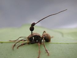
After O. unilateralis causes the mandibles of the ant to close upon the leaf vein, it begins to atrophy the striated muscle by destroying the sarcomere connections in the muscle fibers, as well as reducing the density of mitochondria and sarcoplasmic reticula [6]. The reduction in mitochondria lowers the amount of energy available to the mandible muscles and eliminates the ability for the muscle fibers to relax and contract, resulting in the permanent closure of the mandibles on the leaf vein (known as the death grip). This action is essential in ensuring that O. unilateralis remains at the proper height above the forest floor for the next steps of fungal development and spore release. Within 24 hours of death, hyphae are seen growing from a number of areas on the ant. Hyphae that protrude from the tarsae of the ants function to secure the ant to the leaf. As the hyphae begin to collect and mass together, stroma can be seen forming at the intersegmental membrane at the base of the ant’s head around the 2 day mark. [10]
Fungal reproduction is only possible after a stalk is grown out of the host’s head by propulsion of spores out from its fruiting bodies. Research shows that post mortem stalk growth is adaptive of Ophiocordyceps unilateralis sensu lato, since infected worker ants leave the dry canopy for the humid understory securing a stable microclimate to grow [6].
The fungus’s weapon is the stromatal plate which are produced laterally from its stalk. Inside the stroma are structures called asci which contain spores. The spores are released when there is pressure on the ant cadaver. It is believed that this mechanism is activated at the peak of ant activity where potential targets may be in range [3].
Current Research
In 2011, Hughes et al. studied the behavioral and morphological mechanisms of the fungus O. unilateralis (Hughes et al., 2011). Countless hours of observation of the infected ants' walking patterns resulted in an observed random walking pattern as the research team had predicted, instead of a directed walk (Hughes et al., 2011). While we now have a better understanding than before about the advanced and complex morphological and behavioral mechanisms that happen inside the ant while it is becoming a zombie, there is still much more to know about how the fungus O. unilateralis interacts with ant neurons on the molecular level. Genetic analysis should be done to first understand which proteins of the fungus are interacting with which genes of the ant genome (Hughes et al., 2011). A body of current research on O. unilateralis concerns these valuable interactions which may prove to be useful in human behavioral medicine in the future.
Current research is being done on the possibility that the organism could contain novel chemicals of medicinal use to humans. One of the more interesting recent findings was the presence of anti-malarial naphthoquinones within the organism (citation needed).
Cool Factor
By far the most interesting fact about the organism O. unilateralis is that it has the ability to take the body of an ant captive, turning it into a so-called "zombie ant" and manipulating its psychomotor functions via the release of toxins [2].
The evidence from fossil samples those taken from the Messel Pit in eastern Germany of leaves from Byttnertiopsis daphnogenes when compared to leaves in the Peninsula Khao Chong Botanic Garden in Thailand shows similar bite patterns or “death grip,” of ants infected with Ophiocordyceps unilateralis. The unique bite patterns of the leaves from the Messel Pit were originally believed to be typical vein-cutting behavior common to herbivorous insects. However this hypothesis was discounted when the bite patterns did not match the usual patterns associated with vein-cutting. Instead the bite marks distinctively resembled those of ants infected with O. unilateralis, with the addition that the marks were around major veins. Radiometric dating of the leaves show that they are around 47 million years old, which provides evidence that the parasite manipulation found in O. unilateralis is one of the oldest interactions documented [4].
<iframe width="560" height="315" src="http://www.youtube.com/embed/XuKjBIBBAL8" frameborder="0" allowfullscreen></iframe>
References
1. Van Pelt A: The occurrence of a Cordyceps on the ant Camponotus pennsylvanicus (De Geer) in the Highlands, N.C. region. Journal of the Tennesee Academy of Sciences 1958, 33(120-122).
2. Hughes DP, Evans HC, Hywel-Jones NL, Boomsma JJ, Armitage SAO: Novel fungal disease in complex leaf-cutting ant societies. Ecological Entomology 2009, 34(2):214-220.
3. Evans HC, Samson RA: Cordyceps species and their anamorphs pathogenic on ants (Formicidae) in tropical forest ecosystems. II. The Camponotus (Formicinae) complex. Transactions of the British Mycolocical Society 1984, 82:127-150.
4. Hughes, David P., Torsten Wappler, and Conrad C. Labandiera. "Ancient Death-grip Leaf Scars Reveal Ant-fungal Parasitism." Biology Letters (2010).
5. http://www.physorg.com/news201343671.html
6. Hughes, David P., et al. "Behavioral Mechanisms and Morphological Symptoms of Zombie Ants Dying from Fungal Infection." BMC Ecology 11.1 (2011): 13-22. Print.
7. Wongsa P, Tasanatai K, Watts P, Hywel-Jones N (2005) Isolation and in vitro cultivation of the insect pathogenic fungus Cordyceps unilateralis. Mycol Res 109:936–940.
8. Amnuaykanjanasin A, Phonghanpot S, Sengpanich N, Cheevadhana- rak S, Tanticharoen M (2009) Insect-specific polyketide syn- thases (PKSs), potential PKS-nonribosomal peptide synthetase hybrids, and novel PKS clades in tropical fungi. Appl Environ Microbiol 75(11):3721–3732.
9. . David P. Hughes, et al. "Graveyards On The Move: The Spatio-Temporal Distribution Of Dead Ophiocordyceps-Infected Ants." Plos ONE 4.3 (2009): 1. EDS Foundation Index. Web. 27 Feb. 2012.
10. David P. Hughes, et al. "The Life Of A Dead Ant: The Expression Of An Adaptive Extended Phenotype." The American Naturalist 174.3 (2009): 424-433. JSTOR Life Sciences. Web. 27 Feb. 2012.


