Bovine Herpesvirus 1: Difference between revisions
| Line 27: | Line 27: | ||
==Genome Structure== | ==Genome Structure== | ||
<br> | <br> | ||
Like all herpesviruses, BoHV-1 has a relatively long, double stranded DNA genome. Although packaged linearly, it is thought to circularize during its infectious cycle prior to genome replication. | Like all herpesviruses, BoHV-1 has a relatively long, double stranded DNA genome. Although packaged linearly, it is thought to circularize during its infectious cycle prior to genome replication. It is 135,300 base pairs in length, has a 72% guanine/cytosine content, and has 73 open reading frames (ORFs). Of these 73 ORFs, 33 have been determined to be essential for viral replication. Most of the virus’s genes have homologous counterparts common to all alphaherpesviruses, although some are exclusive to the Varicelloviruses. Only one of these genes (UL0.5) is specific to BoHV-1 alone.<br><br> | ||
BoHV-1 has a class D herpesvirus genome, which means that its genome contains two unique regions, one long and one short (UL and US), the second of which is flanked by inverted repeats (IR and TR). Both of these unique regions can be inverted, meaning that there are actually four isomeric forms of the herpesvirus genome that can be found in a BoHV-1 virion. In reality, one specific orientation of UL is strongly preferred, so only two isomeric forms of the genome are commonly found in the environment (one corresponding to each inversion of US). These inverted sequences are likely attributable to recombination during genome replication. | |||
Most of the virus’s genes | |||
<br> | <br> | ||
Revision as of 06:43, 23 September 2012
A Viral Biorealm page on the family Bovine Herpesvirus 1
Baltimore Classification

Image source: http://www.stdgen.lanl.gov/stdgen/bacteria/hhv2/herpes.html Los Alamos National Laboratory Bioscience Division
Group 1: Double-Stranded DNA Viruses
Higher Order Categories
Order: Herpesvirales
Family: Herpesviridae
Subfamily: Alphaherpesviridae
Genus: Varicellovirus
Description and Significance
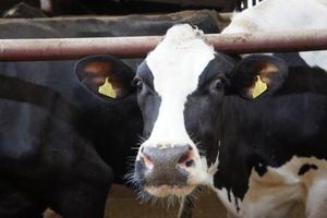
Bovine herpesvirus 1 (BoHV-1) is a well-studied pathogen that infects cattle in many parts of the world. An infected animal can exhibit a wide variety of symptoms depending on the specific conditions of infection, including lesions, respiratory problems, and increased abortion rates. BoHV-1 can be transmitted through various means, including the exchange of nasal discharge, semen transmission, and aerosol transmission. This makes the virus highly contagious, especially in a crowded feedlot environment. BoHV-1 is also capable of entering a latency phase within the host, meaning that an apparently healthy animal might suddenly resume the virus’s spread.
Although infection by BoHV-1 is not usually lethal, it does detract from the productivity of the animal. If the infection is not contained and allowed to spread to the entire herd, there will be significant economic losses incurred from deaths, abortions, reduced body mass, reduced dairy production, and the cost of treatment. Infection by BoHV-1 is also one of the many contributing factors that can lead to the development of the general disease complex Bovine Respiratory Disease (BRD), which is estimated to cost the United States cattle industry about 640 million dollars annually.
Genome Structure
Like all herpesviruses, BoHV-1 has a relatively long, double stranded DNA genome. Although packaged linearly, it is thought to circularize during its infectious cycle prior to genome replication. It is 135,300 base pairs in length, has a 72% guanine/cytosine content, and has 73 open reading frames (ORFs). Of these 73 ORFs, 33 have been determined to be essential for viral replication. Most of the virus’s genes have homologous counterparts common to all alphaherpesviruses, although some are exclusive to the Varicelloviruses. Only one of these genes (UL0.5) is specific to BoHV-1 alone.
BoHV-1 has a class D herpesvirus genome, which means that its genome contains two unique regions, one long and one short (UL and US), the second of which is flanked by inverted repeats (IR and TR). Both of these unique regions can be inverted, meaning that there are actually four isomeric forms of the herpesvirus genome that can be found in a BoHV-1 virion. In reality, one specific orientation of UL is strongly preferred, so only two isomeric forms of the genome are commonly found in the environment (one corresponding to each inversion of US). These inverted sequences are likely attributable to recombination during genome replication.
Virion Structure of Bovine herpesvires 1

Image source: http://www.stdgen.lanl.gov/stdgen/bacteria/hhv2/herpes.html Los Alamos National Laboratory Bioscience Division
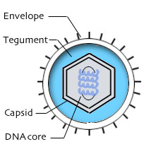
BoHV-1 has a typical herpesvirus virion structure: the virus's double stranded DNA genome is contained within an icosahedral protein capsid. The capsid is wrapped in a protein complex called the tegument, which is made up of about 20 viral proteins. The tegument connects the capsid with the outer cell-derived envelope, which contains the viral membrane proteins and glycoproteins that are essential for the successful penetration of the cell membrane, including glycoproteins gD, gB, gH, and gL.
Reproductive Cycle of Bovine herpesvirus 1 in a Host Cell
The BoHV-1 virion penetrates the cell membrane via a three step process. First, glycoproteins gB and gC on the virion envelope interact with certain cellular structures, creating a low affinity attachment between the virus and the host cell. Second, glycoprotein gD binds to cell membrane protein nectin-1, an immunoglobulin protein. This initiates the third phase, during which the virion envelope fuses with the cell membrane, allowing the capsid and tegument to enter the cytoplasm.
Upon entering the host cell, the virion begins to move towards the nucleus. As it is transported, the tegument proteins surrounding the capsid are shed into the cytoplasm. Although many of these tegument proteins are poorly understood, some are known to have important functions, such as disabling host defenses or subverting the host's resources. For example, the virion host shutoff (vhs) tegument protein is responsible for halting the host's regular protein synthesis, and tegument protein VP16 is required to induce expression of early BoHV-1 genes.
Once it has breached the nuclear membrane, the viral genome is thought to circularize, and a combination of viral and cellular proteins induces the expression of the "immediate early" (IE) genes. The IE gene products induce the expression of "early" genes, at which point viral DNA replication begins. "Early late" gene expression begins during viral genome replication, and then finally the "true late" genes are expressed, which code for the capsid-forming structural proteins.
Meanwhile, replication of the now-circular viral genome proceeds via a "rolling circle" mechanism, which produces multiple genomes connected to each other in sequence from head to tail (concatemeric DNA). This long strand of DNA is then cleaved into individual copies of the viral genome, which are loaded into the newly-formed virion capsids without leaving the nucleus.
How the virion capsid proceeds to exit the nucleus and acquire its tegument proteins and outer envelope is a matter of debate. The leading theory suggests that the mature capsid acquires a primary envelope as it buds out of the inner nuclear membrane, and into the perinuclear space. The new primary membrane then fuses with the outer nuclear membrane, so that the naked capsid is released into the cytoplasm. The capsid then acquires its tegument coating and final envelope by budding with a trans Golgi compartment.
Viral Ecology and Pathology
References
[1] Hage, J.J.; Schukken, Y.H.; Schols, H.; Maris-Veldhuis, M.A.; Rijsewijk, F.A.; Klaassen, C.H. "Transmission of bovine herpesvirus 1 within and between herds on an island with a BHV1 control programme.
[2] Labiuk, Shaunivan L.; Babiuk, Lorne A.; van Drunen Littel-van den Hurk, Sylvia. "Major tegument protein VP8 of bovine herpesvirus 1 is phosphorylated by viral US3 and cellular CK2 protein kinases." J. Gen. Vir. 90 (2009): 2829-39.
[3] Muylkens, Benoit; Thiry, Julien; Kirten, Philippe; Schynts, Frederic; Thiry, Etienne. "Bovine herpesvirus 1 infection and infectious bovine rhinotracheitis." Vet. Res. 38 (2007): 181-209.
[4] Roizman, Bernard; Thayer, Nina. "Herpesvirus Family: Herpesviridae." 2001. http://stdgen.northwestern.edu/stdgen/bacteria/hhv2/herpes.html
[5] Van Oirschot, J.T. "Bovine Herpesvirus 1 in Semen of Bulls and the Risk of Transmission." Vet. Quart. 17 (1995): 29-33.
Page authored for BIOL 375 Virology, September 2010
