Herpesviridae: Viral Cycle, Capsid Transport, and Cancer Treatment: Difference between revisions
From MicrobeWiki, the student-edited microbiology resource
| Line 9: | Line 9: | ||
==Cell Cycle== | ==Cell Cycle== | ||
<br>Include some current research, with at least one figure showing data.<br> | <br>Include some current research, with at least one figure showing data.<br> | ||
[[Image:UL6 portal Cryo-EM.jpg|thumb|300px|right|Cryo-EM tomograpgy of the capsid portal formed by a 12-mer of the UL6 protein. The portal forms at a vertex of the capsid.Cardone et al, 2007. http://www.sciencedirect.com/science/article/pii/S004268220600804X]] | [[Image:UL6 portal Cryo-EM.jpg|thumb|300px|right|Cryo-EM tomograpgy of the capsid portal formed by a 12-mer of the UL6 protein. The portal forms at a vertex of the capsid. Cardone et al, 2007. http://www.sciencedirect.com/science/article/pii/S004268220600804X]] | ||
[[Image:HSV virion assembly.jpg|thumb|300px|right|Diagram of HSV virion assembly. After exiting the nucleus, secondary envelope formation occurs at two separate sites, and is then later joined together.Mettenleiter et al, 2006. http://www.sciencedirect.com/science/article/pii/S1369527406000968]] | [[Image:HSV virion assembly.jpg|thumb|300px|right|Diagram of HSV virion assembly. After exiting the nucleus, secondary envelope formation occurs at two separate sites, and is then later joined together. Mettenleiter et al, 2006. http://www.sciencedirect.com/science/article/pii/S1369527406000968]] | ||
==Section 3== | ==Section 3== | ||
Revision as of 22:39, 22 April 2013
Introduction

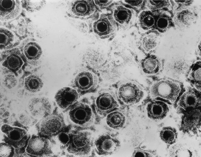
Transmission electron micrograph of herpes simplex virus. Some nucleocapsids are empty, as shown by penetration of electron-dense stain. http://phil.cdc.gov/PHIL_Images/08301998/00014/B82-0474_lores.jpg
By Michael Gallaher
Pathology
Include some current research, with at least one figure showing data.

Cell Cycle
Include some current research, with at least one figure showing data.

Cryo-EM tomograpgy of the capsid portal formed by a 12-mer of the UL6 protein. The portal forms at a vertex of the capsid. Cardone et al, 2007. http://www.sciencedirect.com/science/article/pii/S004268220600804X

Diagram of HSV virion assembly. After exiting the nucleus, secondary envelope formation occurs at two separate sites, and is then later joined together. Mettenleiter et al, 2006. http://www.sciencedirect.com/science/article/pii/S1369527406000968
Section 3
Include some current research, with at least one figure showing data.
Conclusion
Overall text length at least 3,000 words, with at least 3 figures.
References
Edited by student of Joan Slonczewski for BIOL 238 Microbiology, 2011, Kenyon College.
