Neisseria meningitidis causing meningococcal meningitis: Difference between revisions
From MicrobeWiki, the student-edited microbiology resource
| Line 2: | Line 2: | ||
[[Image:N.meningitidis.png|thumb|300px|right|Colored scanning electron micrograph (SEM) of Neisseria meningitidis, gram-negative diplococci that cause meningococcal meningitis (magnified x 33000) (http://www.dailymail.co.uk/sciencetech/article-2197533/As-pretty-picture-lot-deadly--Killer-diseases-youve-seen-before.html).]] | [[Image:N.meningitidis.png|thumb|300px|right|Colored scanning electron micrograph (SEM) of Neisseria meningitidis, gram-negative diplococci that cause meningococcal meningitis (magnified x 33000) (http://www.dailymail.co.uk/sciencetech/article-2197533/As-pretty-picture-lot-deadly--Killer-diseases-youve-seen-before.html).]] | ||
<br>At right is a sample image insertion. It works for any image uploaded anywhere to MicrobeWiki. The insertion code consists of: | <br>At right is a sample image insertion. It works for any image uploaded anywhere to MicrobeWiki. The insertion code consists of: | ||
<br><b>Closed double brackets:</b> ]] | <br><b>Closed double brackets:</b> ]] | ||
<br><br>Other examples: | <br><br>Other examples: | ||
| Line 17: | Line 12: | ||
<br> | <br> <br> | ||
==Section 1== | ==Section 1== | ||
Revision as of 19:56, 24 April 2013
Introduction

Colored scanning electron micrograph (SEM) of Neisseria meningitidis, gram-negative diplococci that cause meningococcal meningitis (magnified x 33000) (http://www.dailymail.co.uk/sciencetech/article-2197533/As-pretty-picture-lot-deadly--Killer-diseases-youve-seen-before.html).
At right is a sample image insertion. It works for any image uploaded anywhere to MicrobeWiki. The insertion code consists of:
Closed double brackets: ]]
Other examples:
Bold
Italic
Subscript: H2O
Superscript: Fe3+
Section 1
Hey there, how are you doing?
Section 2
Include some current research in each topic, with at least one figure showing data.
Section 3
Include some current research in each topic, with at least one figure showing data.
Conclusion
Overall paper length should be 3,000 words, with at least 3 figures.
References
Edited by student of Joan Slonczewski for BIOL 238 Microbiology, 2009, Kenyon College.

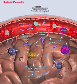
Pathophysiology of Bacterial Meningitis (http://www.qiagen.com/geneglobe/static/images/Pathways/Bacterial%20Meningitis.jpg)..

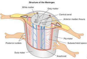
Breakdown of the meninges, a three-layer membrane surrounding the brain and spinal cord (http://body-disease.com/acute-bacterial-meningitis/ ).

Colored scanning electron micrograph (SEM) of Neisseria meningitidis, gram-negative diplococci that cause meningococcal meningitis (magnified x 33000) (http://www.dailymail.co.uk/sciencetech/article-2197533/As-pretty-picture-lot-deadly--Killer-diseases-youve-seen-before.html).
