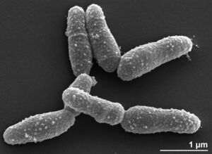
Arcanobacterium haemolyticum
Classification
Bacteria; Actinobacteria; Actinomycetia; Actinomycetales; Actinomycetaceae
Species
|
NCBI: [1] |
Arcanobacterium haemolyticum
Description and Significance
Arcanobacterium haemolyticum is an important human and animal pathogen that is gram-positive and a facultative anaerobe (Vu & Rajnik, 2022). It appears to be a branching bacillus bacteria (Vu & Rajnik, 2022). From swabs and cultures done from swabs, it seems that even in healthy individuals (both human and animal) Arcanobacterium haemolyticum can be found on the skin and pharynx normally (Vu & Rajnik, 2022). As an obligate parasite, Arcanobacterium haemolyticum has been known to cause pharyngitis and skin lesions on some individuals, particularly adolescents and elderly patients (Vu & Rajnik, 2022), which makes it an important microbe to understand.
Genome Structure
The genome of strain 110108T, which is the type strain of Arcanobacterium haemolyticum, is a circular chromosome that is 1,986,154 bp long. 87.2% of the genome is DNA coding (1,744,192 bp), and the GC content of the genome is 53.13% (1,055,308 bp). The genome of Arcanobacterium haemolyticum codes for a total of 1,885 genes, with 64 of those being RNA genes and the other 1,821 genes being protein encoding genes (Yasawong et al., 2010).
Cell Structure, Metabolism and Life Cycle
Arcanobacterium haemolyticum is a Gram-positive, beta-hemolytic bacteria that has two subtypes: rough and smooth (Ruther et al., 2015). The smooth subtype has smooth colony edges, and the rough subtype has rough colony edges (Ruther et al., 2015). Arcanobacterium haemolyticum produces Arcanolysin and phospholipase D which are both virulence factors (Gellings & McGee, 2018). A. haemolyticum is a facultative anaerobe that grows and metabolizes best when it is in the presence of about 5-10% CO2 (Vu & Rajnik, 2022).
Ecology and Pathogenesis
As previously mentioned, Arcanobacterium haemolyticum has been found on the throat as well as on the skin, but the full extent of its habitat is not well understood (Microbe Canvas). Because it is part of the normal microbiome of humans, it can be difficult to determine if it is the pathogen causing the disease in many cases (Vu & Rajnik, 2022). As mentioned before, A. haemolyticum can be classified either by being smooth or rough, with smooth subtypes dominating in wound infections and rough subtypes dominating respiratory infections (Ufkes, 2022). While A. haemolyticum has been isolate in animals, its main host has been proven to be humans (Ufkes, 2022). Though the exact mechanism of infection of cells is not known, it has been proven that A. haemolyticum does have the ability to invade epithelial cells of the upper respiratory tract (HEp-2) and stay there for up to four days which causes an intracellular reservoir of the bacterium (Ufkes, 2022). A couple of important virulence factors produced by A. haemolyticum include phospholipase D which allows cholesterol to be taken in more readily by the host membrane and arcanolysin which interacts with cholesterol which allows the bacteria to form pores in the host cell wall (Ufkes, 2022). It is important to note that A. haemolyticum is thought to be cholesterol dependent as demonstrated by the virulence factors (Ufkes, 2022). The exact pathology and physiology of the rash that sometimes comes with an infection of this sort is not known, yet it is hypothesized to potentially be due to some of the exotoxins released by the bacterium including phospholipase D, neuraminidase, and a hemolysin (Ufkes, 2022). Symptoms of a patient infected with Arcanobacterium haemolyticum can be broad, with some experiencing mild respiratory infection symptoms yet others experiencing severe symptoms that are diphtheria-like (Vu & Rajnik, 2022). Pharyngitis (sore throat) is the most common symptom, and many people will also develop fever, swollen lymph glands, and a rash that spreads from the arms and legs, towards the midline of the body on the neck and back (Vu & Rajnik, 2022). In patients with wound infections, symptoms tend to present as ulcers, abscesses, and cellulitis on the skin (Vu & Rajnik, 2022). In extremely rare cases, A. haemolyticum cases have caused brain abscesses, pneumonia, infective endocarditis, pyothorax, and bacteremia (Vu & Rajnik, 2022).
References
Yasawong, M., Teshima, H., Lapidus, A., Nolan, M., Lucas, S., Glavina Del Rio, T., Tice, H., Cheng, J. F., Bruce, D., Detter, C., Tapia, R., Han, C., Goodwin, L., Pitluck, S., Liolios, K., Ivanova, N., Mavromatis, K., Mikhailova, N., Pati, A., Chen, A., … Klenk, H. P. (2010). Complete genome sequence of Arcanobacterium haemolyticum type strain (11018). Standards in genomic sciences, 3(2), 126–135. https://doi.org/10.4056/sigs.1123072
Vu MLD, Rajnik M. Arcanobacterium Haemolyticum. [Updated 2022 Jul 12]. In: StatPearls [Internet]. Treasure Island (FL): StatPearls Publishing; 2022 Jan-. Available from: https://www.ncbi.nlm.nih.gov/books/NBK560927/
Gellings, P. S., & McGee, D. J. (2018). Arcanobacterium haemolyticum Phospholipase D Enzymatic Activity Promotes the Hemolytic Activity of the Cholesterol-Dependent Cytolysin Arcanolysin. Toxins, 10(6), 213. https://doi.org/10.3390/toxins10060213
Ruther, H., Phillips, K., Ross, D., Crawford, A., Weidner, P., Sammra, O., Lämmler, C., McGee, D. (2015). Smooth and rough biotypes of ARcanobacterium haemolyticum can be genetically distinguished at the arcanolysin locus. PLOS ONE 10(9): e0137346. https://doi.org/10.1371/journal.pone.0137346
https://bacdive.dsmz.de/strain/197
Author
Page authored by Madison Sadler, student of Prof. Bradley Tolar at UNC Wilmington.

