Actinophrydae
A Microbial Biorealm page on the genus Actinophrydae
Classification
Higher order taxa:
Eukaryota, Stramenopiles; Actinophrydae (includes both Actinophrys and Actinophaerium).
Species:
Actinophrys sol, A. vesiculata, A. tauryanini, A. eichhorni
|
NCBI: Taxonomy Genome |
Description and Significance
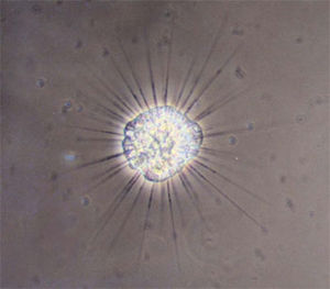
This protist has a spherical body with many pseudopods, called axopodia, that branch out. Actinophrys are part of a group called heliozoa, or "sun animalcule," because their axopodia look like beams of light coming of of a sun. Actinophrys is known to sometimes have more than one nucleus. An organism related to Actinophrys is Actinophaerium, about ten times larger in size. Actinophaerium cells can measure up to 1000 microns but generally average 200 to 300 microns, making it one of the largest heliozoans. Actinophrys is much more common than Actinophaerium.
Genome Structure
There has not been a genome project on Actinophrys or Actinophyaerium.
Cell Structure and Metabolism
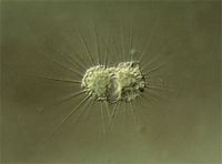
The most noticeable thing about Actinophrys and Actinophaerium are their axipodia.They are attached to the nuclear membrane surface and protrude outwards. The axopodia are supported by axial rods from within the cell. Its axopodia have a number of purposes, capturing or killing prey, and for protection from its predators, which flow along its arms. The axopodia are able to contract quickly to capture their prey. There was a presumed contractile element called contractile tubules structure that was shown to cause the axopodial cytoplasm to create beads along the length of the pseudopod. They are then able to hold onto the prey because of the protein gp40, which is stored in secretory glands called extrusomes. The protein gp40 also contributes to self recognition.
Actinophrys and Actinophaerium obtain energy heterotrophically. The axopodia will capture the prey and bring it to the body, which will then form a food vacuole to surround and digest the organism. After this the organism is lysed, the contents of the vacuole coagulate and much of the fluid is removed from the vacuole. Then the rest of the undigested food in the vacuoles is egested.
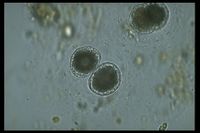
Generally both Actinpophrys and Actinophaerium reproduces asexually through binary fission. But the species Actinophryis heliozoa reproduce sexually through autogamy, or self-fertilization. Actinophryid heliozoa will go through encystment and divide into two diploid cells called gamonts within the cyst. Then both of these cells goes through two meiotic division, but one nucleus in each division becomes degenerated. The two remaining haploid cells fuse together and create a zygote within the cyst. When the environment is suitable the new heliozoan will leave its cyst.
Ecology
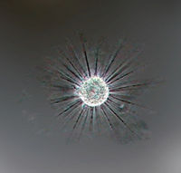
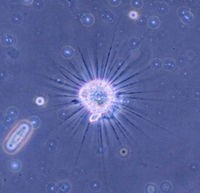
Actinophrys and Actinophaerium are found in open fresh water floating among filamentous algae and reeds. They can be found in large numbers where they are found, but sometimes they can be found singly. They stay in the algae growths to find their prey. Even though they are generally found in open waters like creeks, ponds, lakes ect. they can be found in wet moss. They are a food source for other small protists and invertebrates.
References
Actinophrys "A well-known sun animalcule"
Miscape Magazine. February 2002.
Patterson D.J., Hausmann K. Feeding by Actinophrys sol (Protista, Heliozoa): 1 light microscopy.
Microbios. 1981;31(123):39-55.
Protist Information Server. Protist Images Actinophraerium.
Rotkiewicz, Piotr. Droplet - Microscopy of the Protozoa.
Sakaguchi M., Murakami H., Suzaki T.
The Astrobiology Institute Marine Biological Laboratory. Actinosphaerium eichhornii.
