Introduction

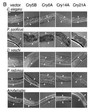
(B) Photographs of the anterior intestine of nematodes fed the four toxic crystal proteins in E. coli. Arrowheads delineate the width of the intestine at one position near the anterior. (Bar 5 20 mm.)<
http://www.pnas.org/content/100/5/2760.long/>

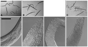
Figure 1. Infection of C. elegans by B. thuringiensis and B. anthracis. Top row: Dissecting microscope view of nematodes cultured under various conditions. Scale bar of all images in top row is 500 mm. Bottom row: Compound microscope view of nematodes cultured under various conditions. For all images in the bottom row, anterior of the worm is top right and scale bar is 50 mm. (A) C. elegans cultured in a well with B. thuringiensis without Cry5B. Top row: None of the six nematodes are infected. All are healthy. The blur associated with some of the worms in the top row is due to their movement in the well. Bottom row: The internal structures of C. elegans fed B. thuringiensis without Cry5B, including the pharynx and intestine, are all intact. (B) C. elegans cultured in a well with B. thuringiensis and Cry5B. Top row: Five of the six worms are completely infected (rigid, lack of internal structures and normal coloration); one is not. Bottom row: Infected animals show complete or near complete digestion of internal structures by the bacteria. Vegetative and sporulated bacteria can be seen in these lethally infected animals. (C) Similar images as in (B) except the bacterium cultured with the nematodes is Bacillus anthracis. <
http://www.plosone.org/article/info%3Adoi%2F10.1371%2Fjournal.pone.0029122/>

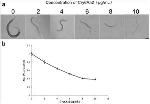
Growth assay of L1 larvae of C. elegans with Cry6Aa2 toxin. a Microscope views of worms cultured in gradient doses of Cry6Aa2 toxin. Scale bar is 100 μm. b The size percentages of worms cultured in a serial dose of Cry6Aa2 toxin to worms cultured in the absence of Cry6Aa2 toxin. Data represent the average of 20 measurements for each toxin concentration. Error bars denote standard deviation <
http://link.springer.com/article/10.1007%2Fs00253-013-5249-3/fulltext.html/>
By Sarah Adrianowycz
At right is a sample image insertion. It works for any image uploaded anywhere to MicrobeWiki. The insertion code consists of:
Double brackets: [[
Filename: PHIL_1181_lores.jpg
Thumbnail status: |thumb|
Pixel size: |300px|
Placement on page: |right|
Legend/credit: (B) Photographs
of the anterior intestine of nematodes fed the four toxic crystal proteins in E. coli. Arrowheads delineate the width of the intestine at one position near the
anterior. (Bar 5 20 mm.)
Closed double brackets: ]]
Other examples:
Bold
Italic
Subscript: H2O
Superscript: Fe3+
Introduce the topic of your paper. What microorganisms are of interest? Habitat? Applications for medicine and/or environment?
Section 1
Include some current research, with at least one figure showing data.


