Leptospira Interrogans
This is the leader section -- here you can write a short description of your topic and why it is interesting. The goal is for this to quickly cover the main ideas of your topic and get the reader interested in reading the rest!
Classification
Higher Order Taxa
Bacteria; Spirochaetes; Spirochaetia; Spirochaetales; Leptospiraceae; Leptospira
Species
Leptospira interrogans
Cell Structure and Metabolism
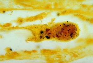
Leptospira interrogans is a gram negative bacteria. The cell is thin and spiral shaped with a hook on each end. It is motile and possesses two periplasmic flagella. The L. interrogans species can be broken down into roughly 250 serovars. Leptospiral serovar diversity results from structural differences in carbohydrate component of lipopolysaccharides. Many serovars are adapted for specific mammalian reservoir hosts. Leptospira interrogans is an aerobic bacteria and possesses both catalase and oxidase enzymes and does not ferment carbohydrates.
Genomic Structure
Mechanisms of Infection
Symptoms and Impact
Humans
Other Mammals
Prevention of Leptospirosis
Due to the common transmission of L. interrogans via rodents, one of the most effective methods of prevention in animals is minimizing rodent populations near animal residences. Pets can also be vaccinated against Leptospirosis, but the vaccine only inhibits a handful of serovars. There is no formal method of Leptospirosis prevention in humans. Chance of contraction can be reduced by minimizing or avoiding direct contact with animal excrement or water contaminated with animal waste.
Current Research
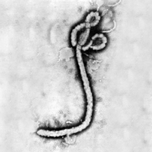
At right is a sample image insertion. It works for any image uploaded anywhere to MicrobeWiki. The insertion code consists of:
Double brackets: [[
Filename: Ebola virus 1.jpeg
Thumbnail status: |thumb|
Pixel size: |300px|
Placement on page: |right|
Legend/credit: Electron micrograph of the Ebola Zaire virus. This was the first photo ever taken of the virus, on 10/13/1976. By Dr. F.A. Murphy, now at U.C. Davis, then at the CDC.
Closed double brackets: ]]
Other examples:
Bold
Italic
Subscript: H2O
Superscript: Fe3+
Overall paper length should be 3,000 words, with at least 3 figures with data.
Further Reading
[Sample link] Ebola Hemorrhagic Fever—Centers for Disease Control and Prevention, Special Pathogens Branch
References
Edited by Emily Gratke, a student of Nora Sullivan in BIOL168L (Microbiology) in The Keck Science Department of the Claremont Colleges Spring 2015.


