Plasmodium vivax
Classification
Domain: Eukaryote
Phylum: Apicomplexa
Class: Aconoidasida
Order: Haemosporida
family: Plasmodiidae
Species
|
NCBI: Taxonomy |
Plasmodium vivax
Description and Significance
Appearance
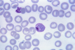
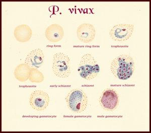
P. vivax are parasites found inhabiting the liver and blood at various stages of development and shape. Starting as rings within red blood cells, then trophozoites as they develop within the red blood cells. Next, they form round gametocytes filling the red blood cells and schizonts which are elongated and wormlike, further filling out the red blood cells. Red blood cells infected by P. vivax cause swelling of the cell, increasing the size by approximately 1.5 times the size.
Habitat
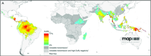
P. vivax is found within South America and Asia, along with smaller amounts found within the horn of africa and Madagascar. P. vivax is the most widespread of human malaria, with farther reaches than the temperate limited Plasmodium falciparum more commonly known, enabled by the parasite forming a dormant stage within the human liver, enabling safe storage for the parasite during mosquito-free cold seasons.[b]
Signifigacne
P. vivax has great signifigance due to the formation of hypnozoites within livers of infected human hosts. This hypnozoite stage leads to a "hybernation" where P. vivax remains dormant until unknown (but linked to mosquito vector bites[b]) factors trigger growth, and relapses of symptoms weeks, months, or years later, remaining infectious throughout [c]. This continual infectious period makes treatment, eradication, and control of the parasite difficult.[c]
Genome Structure
Describe the size and content of the genome. How many chromosomes? Circular or linear? Other interesting features? What is known about its sequence?
P. vivax has a genome characterized by 12-14 linear chromosomes found within a nucleus with a range of 1.2 Mb to 3.5 Mb, with an estimated genome of 35 - 40 Mb. Protein-coding genes have been attributed to GC-rich isochores and chromosome ends with AT-rich isochores at the telomeres.[d]
Cell Structure, Metabolism and Life Cycle
Life Cycle / Cell Structure
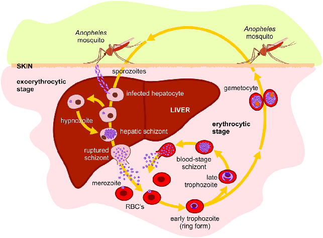
Plasmodium genus eukaryotes form a typical lifecycle characterized by infection from insect host to vertebrate host. P. vivax first enters a vertebrate host through injection by Anopheles mosquitoes (female only) where P. vivax makes their way from the blood to the liver where they , entering hepatic cells and undergoing replication before exiting to infect erythrocytes and forming hypnozoites and merozoites, some differing to form gametocytes to be picked up by other mosquitoes to continue the cycle.
Human Infection
P. vivax starts the infection cycle through an infected mosquito sending saliva into an injection wound while extracting blood to stop coagulation of the blood, sending sporozoites from the mosquito's salivary glands into the blood stream[], reaching the liver through gliding motility[]. Once the sporozoites reach the liver they enter the hepatic cells within and begin to reproduce asexually forming trophozoites and merozoites within.[d]
Liver Stage
The sporozoite enters the hepatocyte and starts multiple rounds of division without segmentation creating a large parasite until segmenting into merozoites that rupture out into the bloodstream. Some remain dormant in a hypnozoite stage for weeks to months until being triggered for growth into the segmented merozoites.
Erythrocytic Stage
P. vivax penetrates young red blood cells (reticulocytes) differing from the more commonly studied P. falciparum which invades erythrocytes. once released P. vivax uses 2 proteins to achieve this (PvRBPP-1 and PvRBP-2) and the Duffy blood group antigens (Fy6) to penetrate the red blood cells. Once inside to synthesize its proteins P. vivax obtains its amino acids through de novo synthesis, import from host plasma, and digestion of host hemoglobin then replicates asexually till the red blood cell ruptures, this swelling forms red blood cells larger in size identifiable through dotting on their surface call Schüffner's dots.
Mosquito Infection
Once a female Anopheles mosquito bites an infecter host, the blood taken up during feed can contain gametocytes (along with other stages of P. vivax) that make their way to the mosquito's stomach, once there the gametocytes form into gametes. Microgametocytes develop daughter nuclei that arrange themselves on the edges of the microgametocyte where cytoplasm develops into thin projections where nuclei enter then break off into male gametes (microgametes). Macrogametocytes develop a cone of reception on one side developing into female gametes (macrogametes)[e].
Fertilization
Male gametes search throughout the stomach for female gametes, once found they enter through the cone fo reception, fusing and forming a zygote through 'anisogamy'. Once formed the zygote becomes vermiform and motile, or an ookinete, penetrating the stomach wall and developing a cyst layer and absorbing nutrients, growing in size, and now an oocyst.
Sporogony
The oocyst continues to divide and create daughter nuclei, developing vacuoles within the cytoplasm within along with cytoplasmic masses. These masses elongate and house the divided daughter nuclei, before the oocyst bursts releasing the sporozoites into the hemolymph of the mosquito. The sporozoites migrate to the salivary glands where they remain till being injected into their new vertebrate host, repeating the cycle.
Ecology and Pathogenesis
Habitat; symbiosis; biogeochemical significance; contributions to environment.
If relevant, how does this organism cause disease? Human, animal, plant hosts? Virulence factors, as well as patient symptoms.
Symbiosis
Symptoms
References
[a] Vogel G. The forgotten malaria. Science. 2013;342(6159):684‐687. doi:10.1126/science.342.6159.684
[c] Am J Trop Med Hyg. 2016 Dec 28; 95(6 Suppl): 15–34. doi: 10.4269/ajtmh.16-0141
[d] J. Carlton The Plasmodium vivax genome sequencing project Trends Parasitol., 19 (2003), pp. 227-231
Author
Page authored by Jonathan Ward, student of Prof. Jay Lennon at IndianaUniversity.
