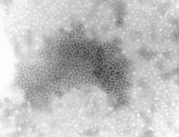
Parvoviridae

Baltimore Classification
Higher order taxa
root; Viruses; ssDNA viruses; Parvoviridae; Parvovirinae
Species
H1 parvovirus, LuIII virus, Raccoon parvovirus (examples)
Description and Significance
Parvoviruses are significant pathogens in the veterinary sciences. These viruses are particularly associated with reproductive failure. A variety of host species are infected by the 3 genera encompassed by the Parvoviridae family. Parvoviruses are among the smallest, simplest eukaryotic viruses and were only discovered in the 1960s while human parvovirus infections were only recognised only in the 1980s. They are naked icosahedral viruses with a genome of a single-strand DNA. The desonucleosis viruses infect insects while the dependoviruses require a helper virus for replication. Although they have been described in a number of mammalian species including man, they are yet to be linked to any disease. The third genus consist of autonomously replicating parvoviruses which are frequently associated with diseases.
Genome Structure
The genome is not segmented and contains a single molecule of linear, negative-sense, single-stranded DNA in most mature virions, or of linear, negative-sense and positive-sense single-stranded DNA in up to 50% in some members. The complete genome has terminally redundant sequences, is repeated at both ends and 5000 nucleotides long. Nucleuotide sequences at the 3'-terminus are complementary to similar regions on the 5' end, or unrelated to the 5'-terminus. The 5'-terminus sequence has palindromic repeats, forming a hairpin structure; terminal repeats at the 5'-end are 200-242 nucleotides long. The 3'-terminus has conserved nucleotide sequences; of 115-116 nucleotides in length; in species of same genus; sequence has hairpin structure. Populations of mature viruses contain particles with equivalent numbers of positive and negative sense ssDNA. The complementary DNA strans usually form dsDNA upon extraction. (source: ICTV dB Descriptions)
Virion Structure of a Parvovirus
The virions of a parpovirus consist of a capsid that is not enveloped and round with icosahedral symmetry. The nucleocapsid is isometric and has a diameter of 20-26nm. The capsid consists of 60 capsomers. Each capsomer is a quadrilateral 'kite-shaped' wedge. The surface projections are small, the surface appears rough and there are distinct spikes.
The capsids can be penetrated by stain and some appear dark in the center. Only one species is recovered in preparations, or the virus may occur together with a dependent virus. (source: ICTV dB Descriptions)
Reproductive Cycle of a Parvovirus in a Host Cell
Comparitively, very little is known about the biology of the pathogenic human parvovirus B 19 because it is difficult to grow in culture. Globotetraosylceramide (Gb4Cer), a glycosphingolipid (erythrocyte P antigen), forms part of the receptor molecule. This explains the cell tropism of B 19.
The nucleus is the site for replication and the scheme above is thought to depict the process. All parvoviruses are highly dependent on cellular functions for gene replication. The cell is required for the autonomous viruses to pass though S-phase for replication to occur but Polyomaviruses cannot turn on host cell DNA synthesis. The defective viruses are even more dependent and require host cell machinery and helper virus for replication.
Very little is currently understood regarding the expression of the Parvovirus genome. The transcription of virus genes is thought to involve the helper function required by the defective viruses. Host cell DNA polymerase is necessary for genome replication.
Dependoviruses can establish a latent infection in the absence of helper virus. The virus genome is integrated into the host cell DNA in this state and can be rescued more than 50 passages lated by Adenovirus infection. The rep gene is involved in this process, but the process is pooly understood. There has been evidence supporting vertical transmission of avian AAV in chickens. At the same time, AAV appears to inhibit cellular transformation by Adenoviruses. (source: Microbiology@Leicester: Parvoviruses)
Viral Ecology & Pathology
The B19 virus is present throughout the year. In temparate climates, outbreaks of infection are more common in the spring and summer. Primary schools are the major targets, where up to 40% of pupils may get infected. 4-10 year olds are most susceptible to infection. 60% of the population are seropositive by adulthood. The usual route of transmission of the virus is respiratory spread. 1 in 40000 blood donations have virus present and bloodborne spread can occur in receipients of whole blood and factor VIII. Haemophiliac children have a significantly higher frequency of seropositivity than is normal.
The B19 virus has been associated with erythema infectiosum, aplastic crisis in patients with chronic haemolytic anaemias, fetal loss in pregnancy and persistent infection in immunocompromised patients. Evidence suporting the presence of B19 in human diseases has already been found. The virus sets up a systemic infection with copious viraemia one week after innoculation. The virus is shed from the respiratory tract at the same time. IgM appears as the viral titres fall and there is a delay of several days before IgG appears.
Reticulocyte members fall to undetectable numbers during viraemia but recover 7 to 10 days later. Normal people lose 1g of Hb. Lymphopenia, neutropenia and thrombocytopenia also occur. The rash appears 17-18 days after innoculation and arthralgia a day or so later: the erythema infectiosum symptoms occur relatively late. These symptoms are likely to be immune mediated. It appears that an early erythrocyte precursor is susceptible to B19 infection. (sources: Virology-Online)
References
Janet Vafaie,MD, and Robert A. Schwartz,MD, MPH; "Parvovirus B19 Infections"; International Journal of Dermatology 2004, 43, 747–749
National Center for Biotechnology Information
