File list
From MicrobeWiki, the student-edited microbiology resource
This special page shows all uploaded files.
| Date | Name | Thumbnail | Size | User | Description | Versions |
|---|---|---|---|---|---|---|
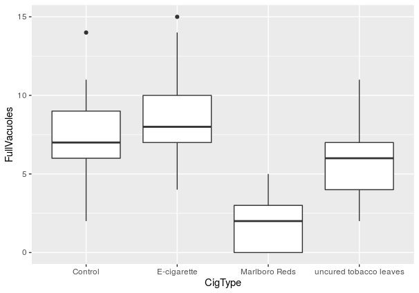
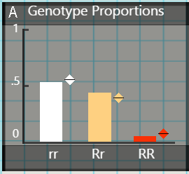
| 00:31, 10 December 2020 | Fragpiechart.png (file) |  |
166 KB | Illuzzi1 | 1 | |
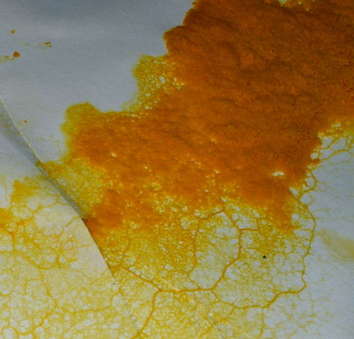
| 00:25, 10 December 2020 | Sclerotium.jpg (file) |  |
43 KB | Beinart1 | 1 | |
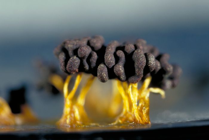
| 23:50, 9 December 2020 | Sporulation.jpg (file) |  |
29 KB | Beinart1 | 1 | |
| 23:45, 9 December 2020 | Genera tree.jpg (file) |  |
52 KB | Mccallie1 | 1 | |
| 22:24, 9 December 2020 | Wiki1.png (file) |  |
62 KB | Burns1 | Figure 1: Showing the TERT gene and the promoter sequence. There is a WT sequence and an Mutated sequence which shows the C>T base pair mutation within. Also it is showing the different Upregulating and Downregulating transcription factors. https://lin... | 5 |
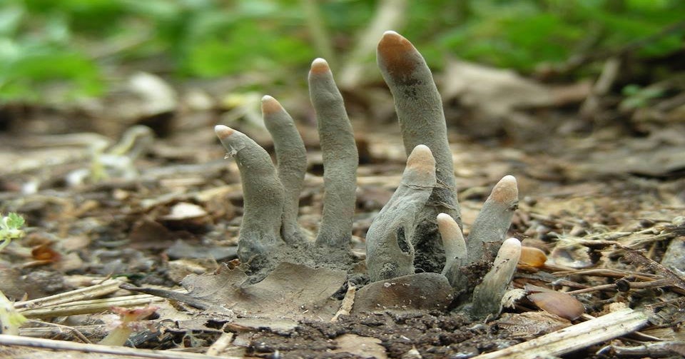
| 19:51, 9 December 2020 | Dead man's fingers fungus.jpg (file) |  |
96 KB | Cjflohrschutz | 1 | |
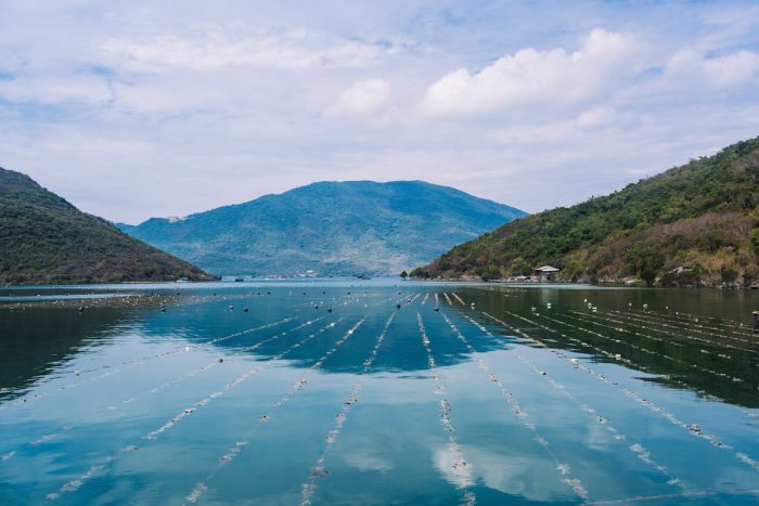
| 19:41, 9 December 2020 | Seaweed.jpg (file) |  |
50 KB | Banthin1 | 1 | |
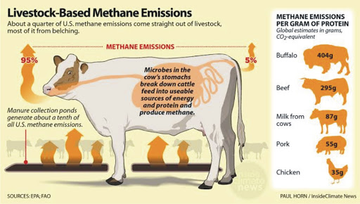
| 19:35, 9 December 2020 | Cow methanogenesis.jpg (file) |  |
53 KB | Banthin1 | 1 | |
| 19:31, 9 December 2020 | CM1 Methanogenesis.jpg (file) |  |
43 KB | Banthin1 | 1 | |
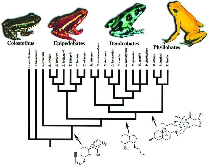
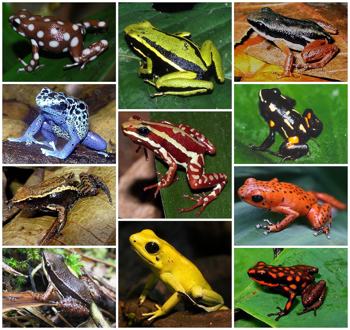
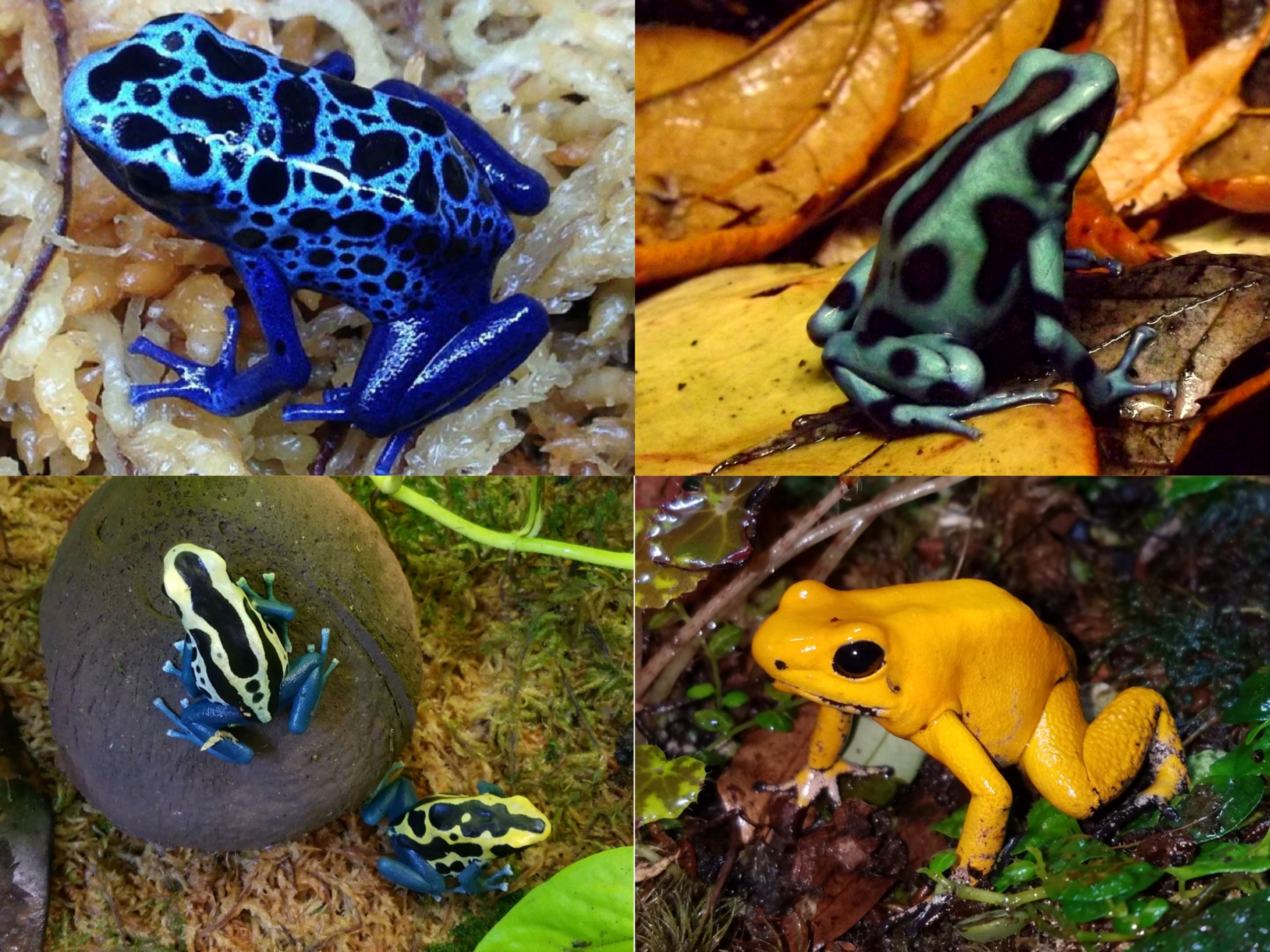
| 19:10, 9 December 2020 | Frog grid.jpg (file) |  |
409 KB | Mccallie1 | 1 | |
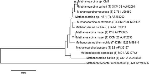
| 18:54, 9 December 2020 | CM1 Tree.jpg (file) |  |
33 KB | Banthin1 | 1 | |
| 16:15, 9 December 2020 | Plasmodial-life-cycle.jpg (file) |  |
52 KB | Beinart1 | 1 | |
| 14:51, 9 December 2020 | 1-plasmodial-slime-mould-nigel-downer.jpg (file) |  |
239 KB | Beinart1 | 1 | |
| 03:59, 9 December 2020 | 4 frogs.jpg (file) |  |
1.19 MB | Mccallie1 | 1 | |
| 03:13, 9 December 2020 | Morphotype.jpg (file) |  |
27 KB | Banthin1 | 1 | |
| 01:49, 9 December 2020 | Moxie.jpeg (file) |  |
10 KB | Pham2 | 1 | |
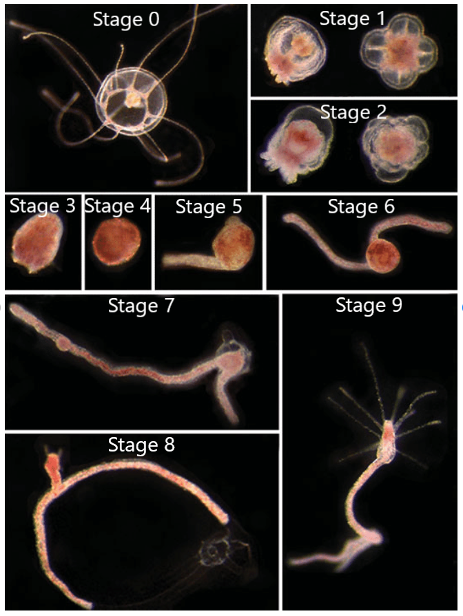
| 01:13, 9 December 2020 | RDStages.png (file) |  |
493 KB | Fitzgerald3 | 2 | |
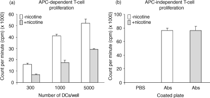
| 23:24, 8 December 2020 | Tcell.jpeg (file) |  |
26 KB | Dougherty1 | 1 | |
| 22:53, 8 December 2020 | Osteoclast.jpg (file) |  |
110 KB | Dougherty1 | 1 | |
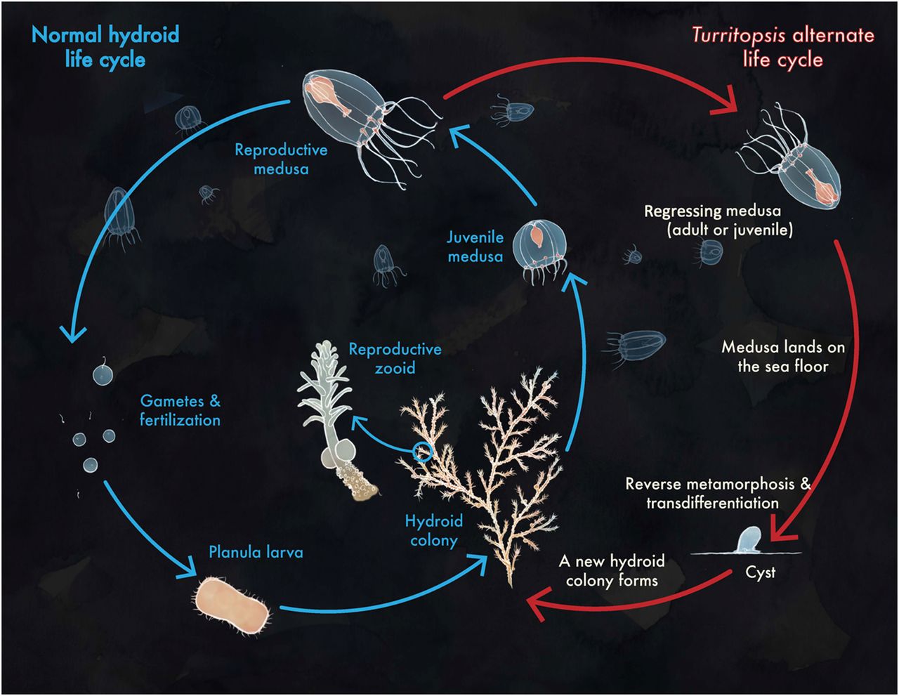
| 20:37, 8 December 2020 | LifeCycle.jpg (file) |  |
141 KB | Fitzgerald3 | 1 | |
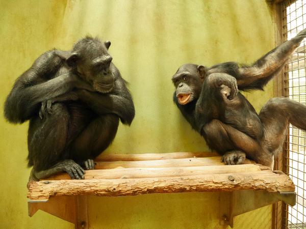
| 20:36, 8 December 2020 | Hirata Kanako.png (file) |  |
153 KB | Schwingel1 | Chimpanzee with Trisomy 22 (right) and euploid chimpanzee (left) interact. Published in 2017 in Hirata et al. "Chimpanzee Down syndrome: a case study of trisomy 22 in a captive chimpanzee"https://link.springer.com/article/10.1007%2Fs10329-017-0597-8 | 1 |
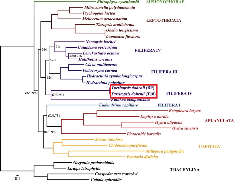
| 20:30, 8 December 2020 | TurritopsisPhylogeny.jpg (file) |  |
124 KB | Fitzgerald3 | 1 | |
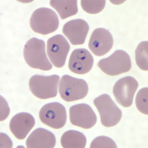
| 17:21, 8 December 2020 | Pf rings thinB.jpg (file) |  |
68 KB | Schwingel1 | Rings of P. falciparum in a thin blood smear https://www.cdc.gov/dpdx/malaria/index.html | 1 |
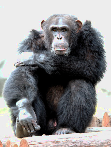
| 17:12, 8 December 2020 | Pan troglodytes .jpg (file) |  |
168 KB | Schwingel1 | Adult male chimpanzee (Pan troglodytes); photo taken 26 October 2006 in Shanghai Wildlife Park, Shanghai, China. Photographer: David Blank https://animaldiversity.org/accounts/Pan_troglodytes/pictures/collections/contributors/david_blank/chimpmale/ | 1 |
| 16:54, 8 December 2020 | FiG3.png (file) |  |
161 KB | Patel2 | 1 | |
| 16:54, 8 December 2020 | FiG2.png (file) |  |
212 KB | Patel2 | 1 | |
| 16:47, 8 December 2020 | FiG1.png (file) |  |
92 KB | Patel2 | 1 | |
| 15:58, 8 December 2020 | Greene fig.3B.png (file) |  |
140 KB | Schwingel1 | Field sample of lances fashioned by chimpanzees for hunting bushbabies in Senegal. Published in H. W. Greene's "Evolutionary Scenarios and Primate Natural History," 2017. Photo credits: J. Pruetz https://www.journals.uchicago.edu/doi/full/10.1086/69283... | 1 |
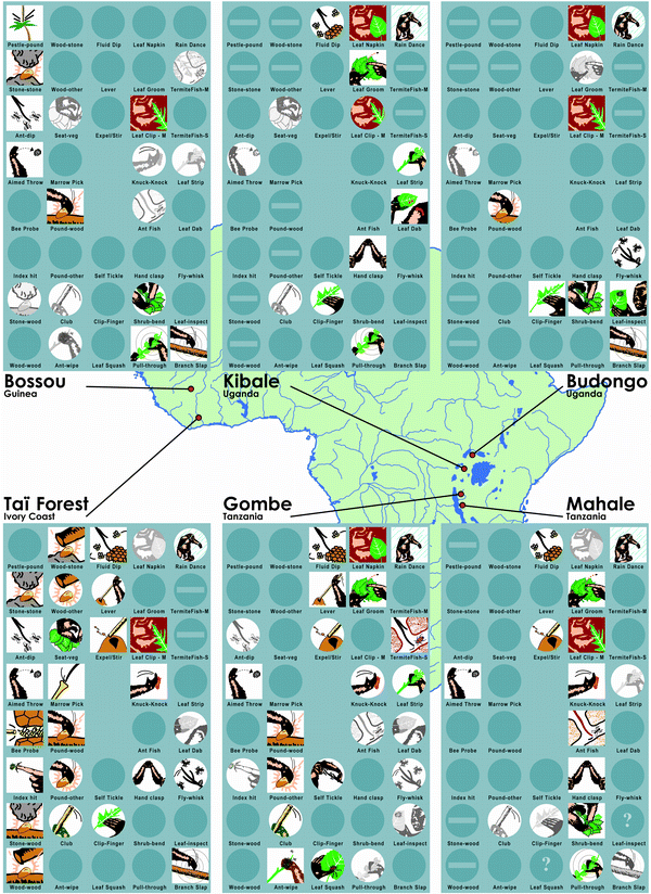
| 15:44, 8 December 2020 | Whiten et al fig.1.png (file) |  |
216 KB | Schwingel1 | Distribution of behavior patterns in chimpanzees across six African study sites. Color icons, customary; circular icons, habitual; monochrome icons, present; clear, absent; horizontal bar, absent with ecological explanation; question mark, answer uncer... | 1 |
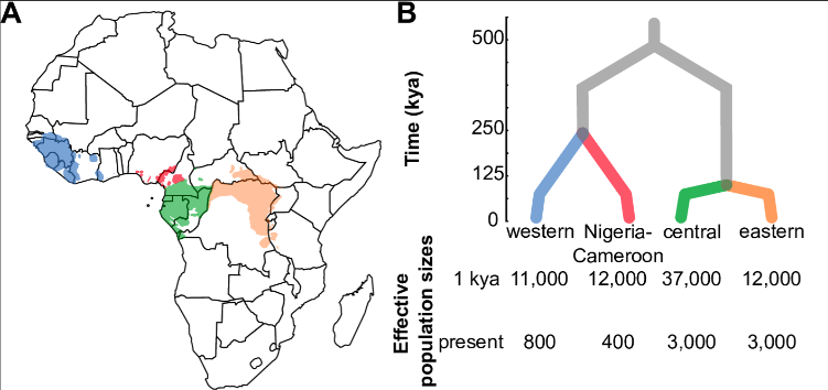
| 15:37, 8 December 2020 | Schmidt Fig. 1 AB.png (file) |  |
85 KB | Schwingel1 | A. The geographic ranges of each chimpanzee subspecies B. Phylogenetic relationships amongst chimpanzees and the timing of their population divergence; 1 kya = long term effective population sizes up to 1kya, present = population sizes 1kya to present.... | 1 |
| 15:08, 8 December 2020 | Greene fig.1.jpg (file) |  |
54 KB | Schwingel1 | Phylogenetic relationships and classification of primates from Harry W. Greene's article "Evolutionary Scenarios and Primate Natural History," published in the American Naturalist, August 2017 | 1 |
| 03:31, 8 December 2020 | Tetrahymena.png (file) |  |
7 KB | Dougherty1 | 1 | |
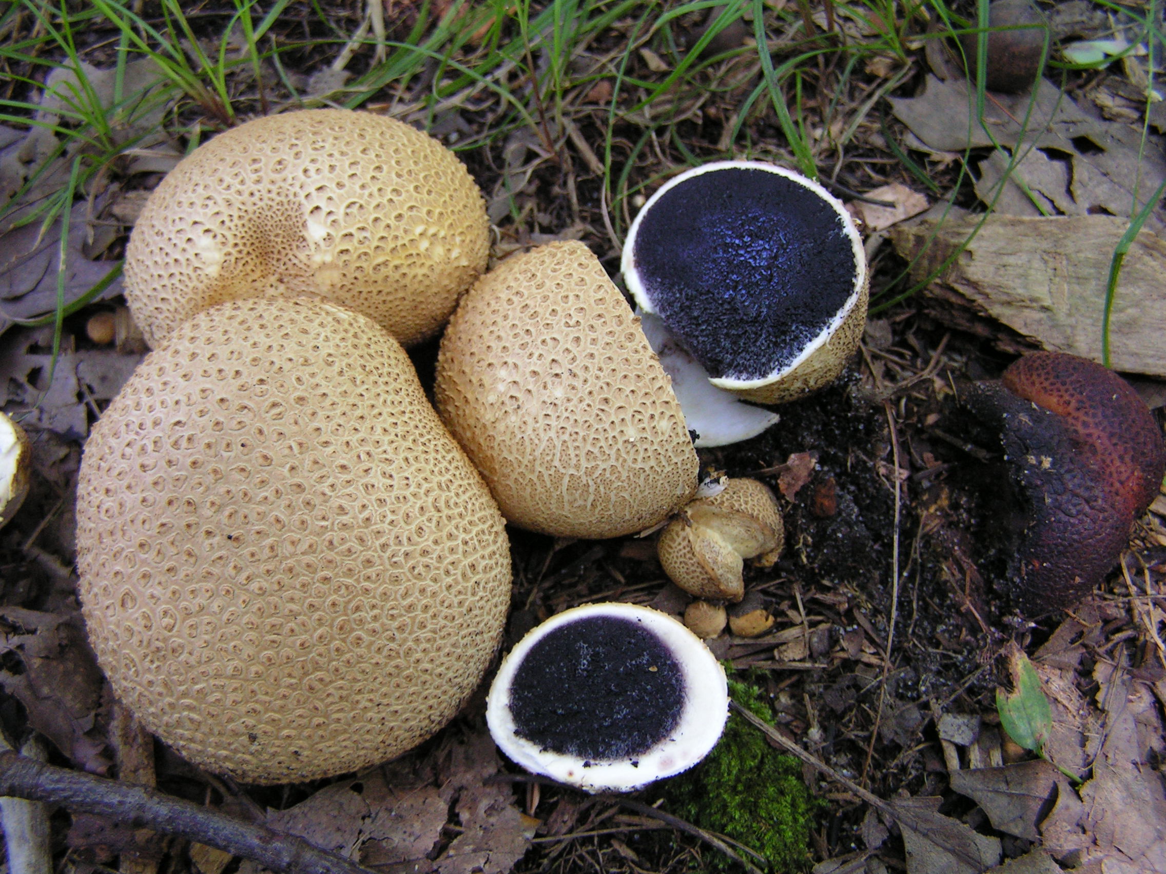
| 00:21, 8 December 2020 | 2009-09-03 Scleroderma citrinum Pers 55647.jpeg (file) |  |
756 KB | Ggourdet | 1 | |
| 19:48, 7 December 2020 | Nicotinebacteria.png (file) |  |
180 KB | Dougherty1 | 1 | |
| 19:46, 7 December 2020 | Nicotinebacteria.webp (file) |  |
60 KB | Dougherty1 | 1 | |
| 18:39, 7 December 2020 | Ant-e1492044072290.jpg (file) |  |
63 KB | Dunst1 | 1 | |
| 18:24, 7 December 2020 | Conifer resin from Agathis lanceolata (New Caledonia, southwestern Pacific Basin) 5 (39352788962).jpg (file) | _5_(39352788962).jpg) |
112 KB | Dunst1 | 1 | |
| 17:17, 7 December 2020 | A tubi .png (file) |  |
436 KB | Raship | The image has been reprinted from [https://commons.wikimedia.org/wiki/File:Aspergillus_tubingensis_FJBJ11.png Qing-Wei Tan, Fang-Luan Gao, Fu-Rong Wang, and Qi-Jian Chen], licensed under the [https://creativecommons.org/licenses/by/4.0/deed.en Creative... | 1 |
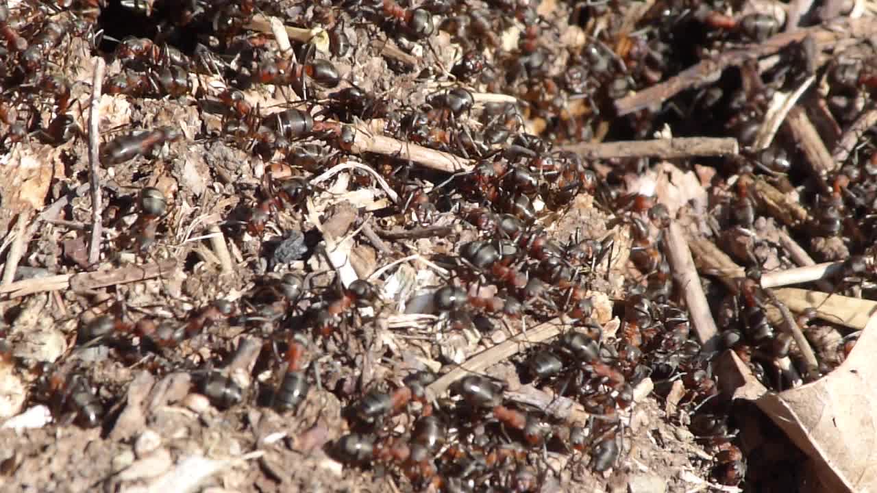
| 17:04, 7 December 2020 | Wood ant (Formica lugubris) (33876593612).jpg (file) | _(33876593612).jpg) |
108 KB | Dunst1 | 1 | |
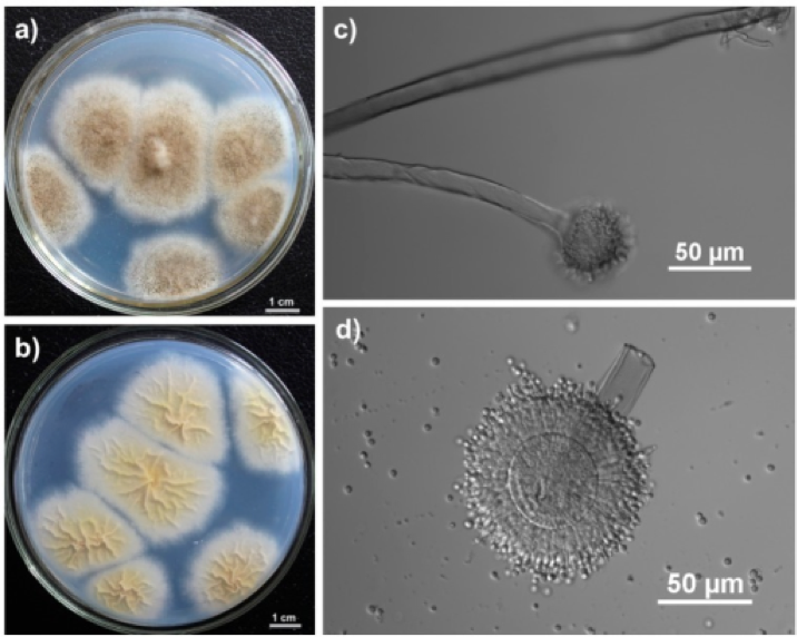
| 17:00, 7 December 2020 | A tubingensis .png (file) |  |
436 KB | Raship | The image has been reprinted from [https://commons.wikimedia.org/wiki/File:Aspergillus_tubingensis_FJBJ11.png Qing-Wei Tan, Fang-Luan Gao, Fu-Rong Wang, and Qi-Jian Chen], licesned under the [https://creativecommons.org/licenses/by/4.0/deed.en Creative... | 1 |
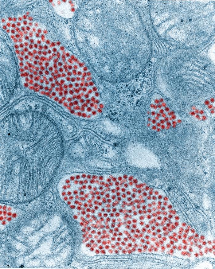
| 15:17, 7 December 2020 | Image0 (2).jpeg (file) | .jpeg) |
241 KB | Ocnieves | This 83,900X magnified image shows a salivary gland tissue section extracted from a eastern equine encephalitis (EEE) virus infected mosquito. This image was taken using transmission electron microscopy, and the EEE virus particles are digitally colori... | 1 |
| 00:32, 7 December 2020 | LifeCycle.png (file) |  |
50 KB | Fitzgerald3 | 1 | |
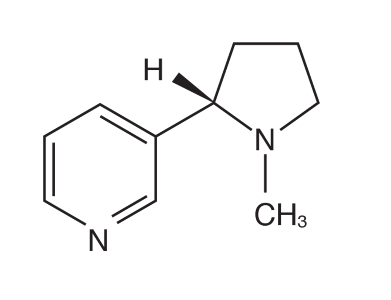
| 00:10, 7 December 2020 | Nicotine.png (file) |  |
73 KB | Dougherty1 | 1 | |
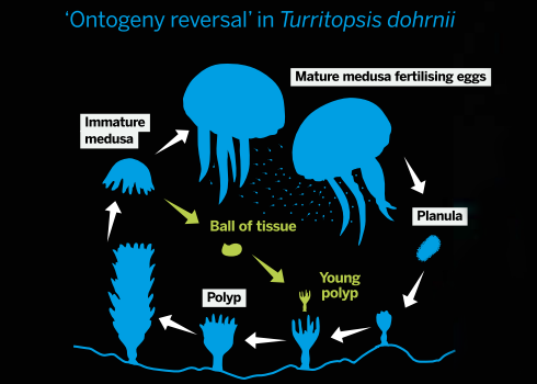
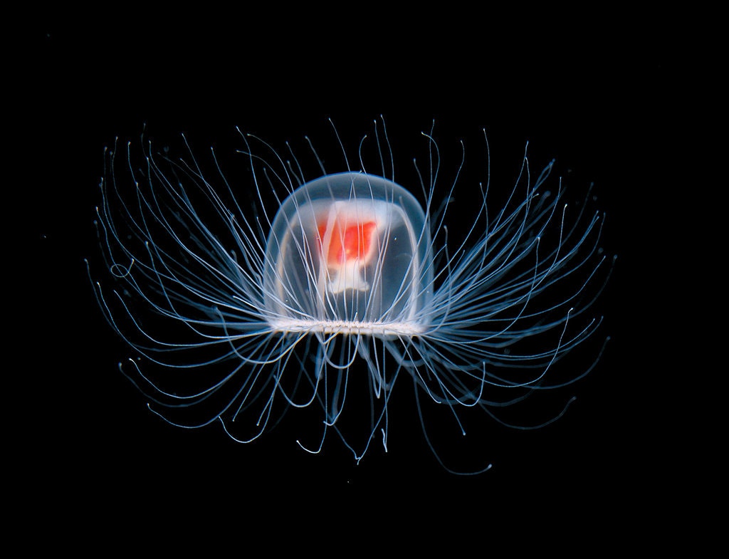
| 21:23, 6 December 2020 | Jellyfish.jpg (file) |  |
100 KB | Fitzgerald3 | 1 | |
| 19:11, 6 December 2020 | Armillaria.png (file) |  |
540 KB | Jmtalbot | 1 | |
| 18:10, 5 December 2020 | The-Largest-Bacteria-in-the-World-2.gif (file) |  |
59 KB | Barrabee1 | 1 | |
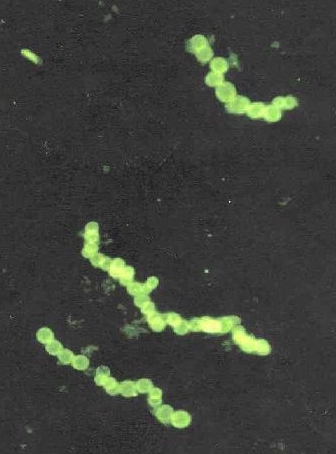
| 18:57, 30 November 2020 | Sulphide bacteria crop.jpg (file) |  |
90 KB | Barrabee1 | 1 | |
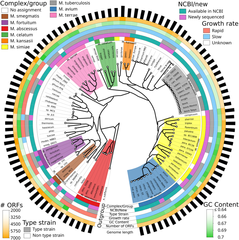
| 22:06, 25 November 2020 | Srep45258-f1.jpg (file) |  |
580 KB | Cebarry | Figure 1. Whole-genome phylogeny of the Mycobacterium genus. Colored blocks of the innermost circle represent Mycobacteria groups based on the phylogeny. M. immunogenum is categorized under the M. abscessus complex. (Retrieved from Fedrizzi et al., 2017) | 1 |
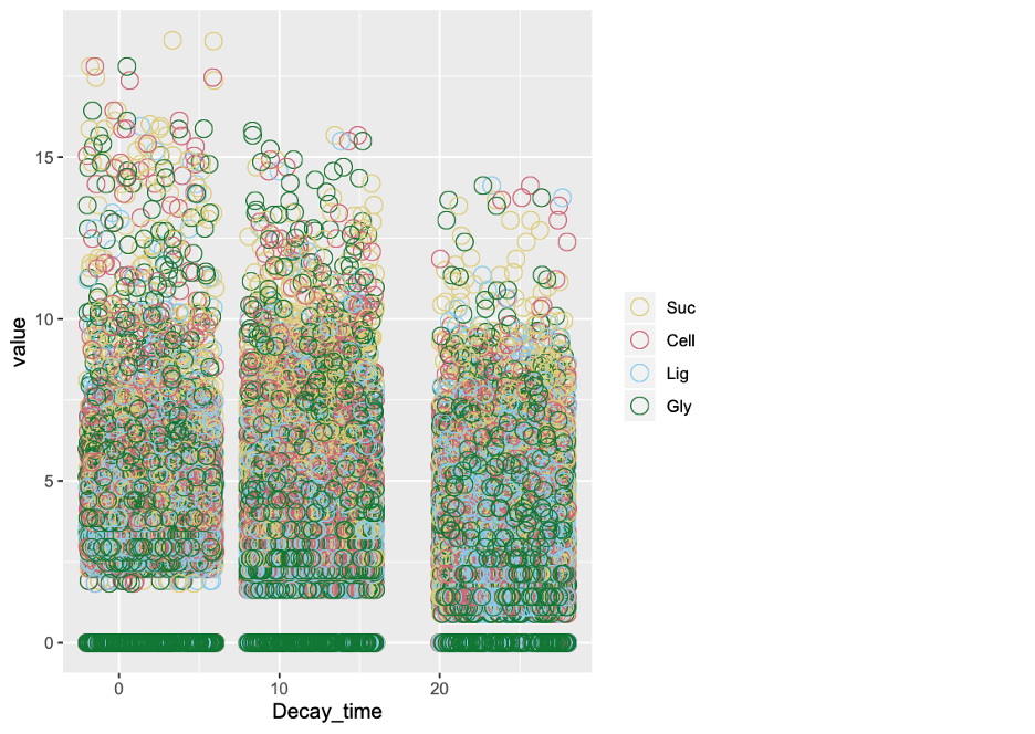
| 21:15, 18 November 2020 | Fish 1.png (file) |  |
14 KB | Slonczewski | Fish data | 1 |
| 16:18, 8 November 2020 | 10815 lores.jpg (file) |  |
59 KB | Slonczewski | CDC by Frederick A. Murphy. Colorized transmission electron microscopic image showing the filamentous and curved morphology of an Ebola virus particle. See PHIL 1181 for a black and white version of this image. | 1 |
