Tuberculosis and HIV Co-Infection
Introduction
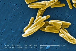
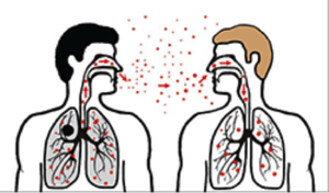
The bacteria Mycobacterium tuberculosis causes tuberculosis. Mycobacterium tuberculosis is an acid fast, rod shaped, gram-positive bacteria that has a unique cell wall compared to other gram-positive bacteria. The cell wall of these bacteria contains mycolic acids, lipid complexes and porins to transfer materials in and out of the cell. In addition to the cell wall these bacteria do not have an outer cell membrane and have layers of arabinogalactan and peptidoglycan under the cell wall. The structure of the unique mycolic acids located in the cell wall make it difficult for nutrients to move in and out of the cell, which results in a slow growth rate compared to other bacteria (Microbewiki, 2012). M. tuberculosis mainly infects the lungs but also has the ability to spread to other areas of the body. One of the reasons that M. tuberculosis usually infects the lungs because M.tubercuolsis is an obligate aerobic and grows best under high oxygen conditions. Mycobacterium tuberculosis is transferred through the air when someone a person who is infected with TB coughs or sneezes and another person inhales the contaminated air. (Tuberculosis, 2014). The tuberculosis infection can exist within the body two in different forms. A Tuberculosis infection is characterized as a latent infection when the body is infected by Mycobacterium tuberculosis and the body’s immune system fights the infection and is able to successfully stop the pathogen from reproducing, growing and spreading. Individuals that have the latent form of TB do not have any symptoms, do not appear to be sick, and cannot transmit the Mycobacterium tuberculosis bacteria to others. A latent TB infection has the ability to become active within the body when the immune system can no longer control the infection. An individual becomes infected with active TB when the immune system cannot stop the bacteria from multiplying inside the body. Symptoms of an active TB infection include chest pain, coughing up blood, fever, fatigue, and weight loss. Some people who come into contact with M. tuberculosis will develop an active TB infection and show symptoms quickly, while it can remain latent in others for years. In some individuals the infection will remain latent and never become active. The development of a TB infection from latent to active is a result of the immune system response of the host. Some individuals are at greater risk for developing active TB infections then others. Individuals who have with compromised immune systems or diseases that make in difficult to fight infection are at a higher risk for tuberculosis. Individuals previously had a TB infection did not get proper treatment are also at a higher risk (Tuberculosis, 2014).
By Michelle Picard
At right is a sample image insertion. It works for any image uploaded anywhere to MicrobeWiki. The insertion code consists of:
Double brackets: [[
Filename: PHIL_1181_lores.jpg
Thumbnail status: |thumb|
Pixel size: |300px|
Placement on page: |right|
Legend/credit: Electron micrograph of the Ebola Zaire virus. This was the first photo ever taken of the virus, on 10/13/1976. By Dr. F.A. Murphy, now at U.C. Davis, then at the CDC.
Closed double brackets: ]]
Other examples:
Bold
Italic
Subscript: H2O
Superscript: Fe3+
By Student Name
At right is a sample image insertion. It works for any image uploaded anywhere to MicrobeWiki. The insertion code consists of:
Double brackets: [[
Filename: PHIL_1181_lores.jpg
Thumbnail status: |thumb|
Pixel size: |300px|
Placement on page: |right|
Legend/credit: Electron micrograph of the Ebola Zaire virus. This was the first photo ever taken of the virus, on 10/13/1976. By Dr. F.A. Murphy, now at U.C. Davis, then at the CDC.
Closed double brackets: ]]
Other examples:
Bold
Italic
Subscript: H2O
Superscript: Fe3+
Introduce the topic of your paper. What microorganisms are of interest? Habitat? Applications for medicine and/or environment?
