Ebola virus entry into host cells
Introduction
Ebola viruses belong to the family, Filoviridae. Ebolavirus is one of two filovire geneses, Marburgvirus and Cuevavirus. There are five known species of ebolavirus; Bundibugyo ebolavirus (BDBV), Zaire ebolavirus (EBOV), Reston ebolavirus (RESTV), Sudan ebolavirus (SUDV), and Taï Forest ebolavirus (TAFV). The virus got its name from the Ebola river, the river that runs near the first discovery site in sub-Saharan Africa. Species such as Zaire ebolavirus (EBOV) cause hemorrhagic fever and have up to a 90% mortality rate in humans (Leroy et al. 2001). Due to the very high mortality rate, this virus is of great concern to scientists today and is considered to be a risk group 4 pathogen. Even species such as Reston ebolavirus, which causes illness in infected, non-human primates but not in humans, are still of great concern because there is a chance that the virus could mutate and become pathogenic to humans (Weingartl et al. 2012). Moreover, since there is no effective vaccine, treatment, or cure for the virus, the virus could potentially be used as a bioterrorism agent. For these reasons, access to the virus is strictly regulated.
The natural reservoir of the virus is still unknown. The virus is believed to have originated in fruit bats, but that has not been confirmed. Outbreaks of ebolavirus tend to occur in places where the consumption of “bushmeat” is commonplace, such as Sub-Saharan Africa. This is strong evidence that the virus infects the human population first through animal-human contact, then through human-human contact.
By Student Name
At right is a sample image insertion. It works for any image uploaded anywhere to MicrobeWiki. The insertion code consists of:
Double brackets: [[
Filename: PHIL_1181_lores.jpg
Thumbnail status: |thumb|
Pixel size: |300px|
Placement on page: |right|
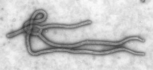
Legend/credit: Electron micrograph of the Ebola Zaire virus. This was the first photo ever taken of the virus, on 10/13/1976. By Dr. F.A. Murphy, now at U.C. Davis, then at the CDC.
Closed double brackets: ]]
Other examples:
Bold
Italic
Subscript: H2O
Superscript: Fe3+
Transmission
By Student Name
At right is a sample image insertion. It works for any image uploaded anywhere to MicrobeWiki. The insertion code consists of:
Double brackets: [[
Filename: PHIL_1181_lores.jpg
Thumbnail status: |thumb|
Pixel size: |300px|
Placement on page: |right|
Legend/credit: Electron micrograph of the Ebola Zaire virus. This was the first photo ever taken of the virus, on 10/13/1976. By Dr. F.A. Murphy, now at U.C. Davis, then at the CDC.
Closed double brackets: ]]
Other examples:
Bold
Italic
Subscript: H2O
Superscript: Fe3+
[[ ]]
Introduce the topic of your paper. What microorganisms are of interest? Habitat? Applications for medicine and/or environment?


