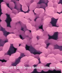
Streptococcus mutans

Classification

Higher order taxa
Bacteria(Domain); Firmicutes(Phylum); Bacilli(Class); Lactobacillales(Order); Streptococcaceae(Family) (2).
Species
Streptococcus mutans (2).
Description and significance
Streptococcus mutans is a Gram-positive bacterium that lives in the mouth. It can thrive in temperature ranging from 18-40 degrees Celsius. The bacterium metabolizes different kinds of carbohydrates, creating an acidic environment in the mouth as a result of this process. This acidic environment in the mouth is what causes the tooth decay. It is the leading cause of dental caries (tooth decay) worldwide. S. mutans is considered to be the most cariogenic of all of the oral Streptococci (8). S. mutans was first described by James Kilian Clarke (1886-1950) after he isolated it from a carious lesion, but it was not until 1960s that real interest in this microbe was generated when researchers began studying dental caries (8).
S. mutans is very important to study, not only because virtually everyone in the world carries it, but also because it has various symptoms that affect our daily lives. As the bacteria develop in the mouth, they cause tooth destruction, impaired speech, difficulty chewing, multiple infections, psychological problems such as low self-esteem, poor social interactions, concentration problems, etc. Though not fatal, tooth decay is one of the most common infectious diseases in humans. Also, cavities caused by the bacteria are the reason for half of all dental visits in the U.S. (6).
Genome structure
The genome of S. mutans UA159, a serotype c strain, has been completely sequenced and is composed of 2,030,936 base pairs. It contains 1,963 open reading frames, 63% of which have been assigned putative functions. Almost 300 appear to be unique to S. mutans. Previously, only three genes for glucan-binding proteins have been isolated, but genome sequencing has uncovered a potential fourth gene, gbpD. Genes associated with transport system are account for almost 15% of the genome. Virulence genes associated with extracellular adherent glucan production, adhesins, acid tolerance, proteases, and putative hemolysins have been identified. Strain UA159 is naturally competent and contains all of the genes essential for competence and quorum sensing. There are no bacteriophage genomes present in S. mutans (7).
S. mutans is composed of circular DNA, and has at least three closely related, but different plasmids. The size of these plasmids are similar, approximately 5.6 kilobase(kb). These plasmids are important to S. mutans because of their functions including resistance to certain anti-biotics or heavy metals, bacteriocin production and immunity, accessory catabolic pathways and mechanisms for conjugation-like transfer activities (4).
Cell structure and metabolism
Streptococcus mutans is a Gram-positive bacteria, has a thick cell wall, and retains a gentian violet. The cell wall is composed of peptidoglycan (murein) and teichoic acids that prevent osmotic lysis of cell protoplast and confer rigidity and shape on cell. S. mutans has a capsule that is composed of polysaccharide, and its structural subunit is dextran glucose. One of the virulence factors of S. mutans in cariogenicity is its ability to attach to the tooth surface and form a biofilm (11). S. mutans attaches to the surface, produces slime, divides and produces microcolonies within the slime layer, and constructs a biofilm. It adheres specifically to the pellicle of the tooth by means of a protein on the cell surface. S. mutans grows and synthesizes a dextran capsule which binds them to the enamel and forms a biofilm of some 300-500 cells. In the metabolism of S. mutans, it is able to cleave sucrose (after consuming carbohydrates provided by the animal diet) into glucose plus fructose. The fructose is fermented as an energy source for bacterial growth. The glucose is polymerized into an extracellular dextran polymer that cements S. mutansto tooth enamel and becomes the matrix of dental plaque. The dextran slime can be depolymerized to glucose for use as a carbon source, resulting in production of lactic acid within the biofilm (plaque) that decalcifies the enamel and leads to dental caries or bacterial infection of the tooth (10).
Ecology
The association of S. mutans in dense biofilms on the teeth suggests that S. mutans may affect other plaque bacteria in the mouth. In fact, earlier studies have shown that there is an inverse relationship between the quantity of S. mutans in dental plague and the presence of another bacterim named Streptococcus sanguinis. The study shows that S. mutans antagonize the growth of S. sanguinis via either acid production or the elaboration of bacteriocins (3).
Pathology
Streptococcus mutans is an animal parasite, especially for animals that have a high carbohydrate (sucrose, fructose, and glucose)diet and is well known as primitive causative agent of dental caries in humans (5).
S. mutans is the main contributor to tooth decay, and is mostly found on surfaces of teeth. One tooth may have a large number of these bacteria, while the tooth next to it may have only a small number. The bacteria are most concentrated in the crevices, pits, and fissures that are a normal part of the teeth and surrounding structures. Adults may have a high concentration of S. mutans in their mouths. In contrast, infants and children have a smaller concentration, but they are more vulnerable to the bacteria (6). S. mutans can be transmitted from a parent or another intimate caregiver to an infant or child via saliva, for example, by allowing infants or children to put their fingers in the parent’s mouth and then into their own mouths, testing the temperature of a bottle with the mouth, sharing forks and spoons, and “cleaning” a pacifier or a bottle nipple that has fallen by sucking on it before giving it back to the infant or child.
Streptococcus mutans has been strongly implicated as the principal etiological agent in dental caries. One of the important virulence properties of these organisms is their ability to form biofilms known as dental plaque on tooth surfaces. Dental plaque formation on tooth surfaces involves three distinct steps. First, formation of the conditioning film or acquired pellicle on the tooth enamel, second, subsequent cell-to-surface attachment of the primary colonizers, and third, cell-to-cell interactions of late colonizers with one another as well as with the primary colonizers (13). Biofilms are sessile bacterial communities adherent to a surface, and their formation occurs in response to a variety of environmental cues. S. mutans undergoes a developmental program in response to environmental signals that leads to the expression of new phenotypes that distinguish these sessile cells from planktonic cells (13). The importance with respect to medicine, biofilm cells have been shown to be up to 1,000-fold more tolerant of antibiotics, and this makes it hard to treat S. mutans with modern medicine.
When food containing carbohydrates is consumed, S. mutans interacts with it and produces acids that cause mineral loss in teeth. The tooth cavity is the result of this mineral loss and can eventually destroy the whole tooth. Tooth decay can spread in the mouth, and can cause extreme pain with difficulty of chewing. Several infections caused by S. mutans can even result a death in extreme cases (6).
Some virulence factors of S. mutans are found that distinguish S. mutans strains from other oral streptococci isolated from the human oral cavity. First, S. mutans is able to synthesize insoluble adhesive glucans from sucorse. Second, it has relatively more acid tolerance (aciduricity). Third, it has a rapid production of lactic acid from dietary sugars. Also, a number of genes that influence the virulence are found. These genes include gtfB, gtfC, and gtfD genes coding for glucosyltransferases, the gbpA and gbpC genes encoding glucan-binding proteins, spaP expressing a cell surface adhesion, and the glgR gene involved in intracellular polysaccharide storage. In addition, a number of other genes that have been shown to affect potential virulence properties in vitro are also characterized, including some involved in the stress responses of S. mutans. These genes are ffh, dgk, gbpB, and an apurinic-apyrimidinic endonuclease gene (3).
Application to Biotechnology
S. mutans does not produce any useful compounds or enzymes that are known in current studies. However, recent studies have found that there are at least 300 genes that are unique to S. mutans, and these are very significant because these are the potential drug targets. Disrupting them would disable the pathogen without harming other bacteria in the mouth (7).
Current Research
In a current study conducted by a group of biologists from University of North Carolina and Washington University found that S. mutans ftf expressionwas affected by both the specific carbohydrate consumed and the age of the host animal. The fructosyltransferase gene (ftf) of S. mutans is a gene that directly associated with infection in the mouth (9). The ftf gene of S. mutans encodes the product fructosyltransferase, a enzyme that catalyzes the cleavage of sucrose, with subsequent polymerization of the fructose moieties into fructan, allowing it to remain in the dental plaque after its production (9). First, they fed animals with different types of carbohydraes: fructose, glucose and sucrose. Then the bacteria activity was observed. As a result, there was the most bacterial activity found on sucrose, and activities on glucose and fructose were similar, which were only 1/3 of bacteria on sucrose (9). Second, animal age was examined as a potential factor of bacteria gene expression. In the result, young animals had been demonstrated to be more susceptible to the formation of dental caries than older animals. This study demonstrated animal age-dependent expression of an S. mutans virulence-associated gene whose product played an important role in the causation of dental caries (9).
Recently, researchers studied the role of WapA gene in S. mutans in cell surface structures, related functions, biofilm formation. Biofilm formation is one of the well-recognized virulence factors of S. mutans, which involves a sucrose independent initial attachment, cell–cell aggregation, a sucrose-dependent stabilization, and eventual biofilm muturarion (11). In order to study the role of WapA in cell surface structures and related functions, they constructed an isogenic mutant of WapA by insertional inactivation, then compared with wild type WapA gene(11). First, they labeled the wild type strain and mutant, and analyzed the architecture of the biofilm in order to see if the morphological change would affect the biofilm architecture. The result showed that the wild type cells fromed a biofilm with large but sparsely distributed microcolonies and large areas occupied by unstructured or single-cell chains. However, the mutant formed a thin biofilm and attached to the surface mostly as unstructured cell layers. Second, they tried to find the effect of WapA mutation on cell surface stickiness of S. mutans. As they immobilized the two cells, they applied force to cell walls to observe the rupture event due to breakage of adhesive bond of each cell. The result showed that wild type cell was stickier than the mutant. Lastly, they tested both wild type and mutant to see an effect of sucrose on gene expression. They measured the gene expression by PCR in absence and presence of sucrose. The result showed that both WapA wildtype and mutant gene expression were repressed by sucrose. The overall results suggested that the WapA protein plays an important structural role on the cell surface, which ultimately affects sucrose-independent cell–cell aggregation and biofilm architecture (11).
A recent study from the University of Heidelberg conducting by Dr. Geiss suggested that titanium, gold, natural enamel and amalgam alloy were superior to composite materials in reducing the adherence of Streptococcus mutans to dental restorations. In the in vitro study, 73 restorative material specimens were coated with cultured S. mutans and then examined with a scanning electron microscope for bacterial adherence (12). Dr. Geiss reported that in the absence of saliva, samples of titanium, gold, natural enamel and amalgam alloy had significantly less S. mutans adherence than Herculite XRV, a composite material manufactured by Kerr Dental (12). However, in the presence of saliva, bacterial adherence of S. mutans was reduced on the titanium and amalgam samples but increased in most of the other tested materials (12). This study also found that the least S. mutans adhered to specimens of titanium, a reactive metal that forms a passivating oxide layer. This findings were very significant in dentistry because Titanium and titanium alloys were commonly used in dental implants and prostheses, crown fabrications, bridge frameworks, and denture frameworks (12).
References
1. Dennis Kunkel Microscopy. http://education.denniskunkel.com/catalog/product_info.php?products_id=9571
2. National Center for Biotechnology Information. http://www.ncbi.nlm.nih.gov/Taxonomy/Browser/wwwtax.cgi?mode=Info&id=1309&lvl=3&lin=f&keep=1&srchmode=1&unlock
3. Vincent A. Fischetti, Richard P. Novick, Joseph J. Ferretti, Daniel A. Portnoy, and Julian I. Rood. "Gram-Positive Pathogens." 2nd ed. Washington: American Society for Microbiology press, 2006
4. Shigeyuki Hamada, Suzanne M. Michalek, et al. "Molecular Microbiology and Immunobiology of Streptococcus mutans." New York: Elsevier Science, 1986
5. Infection and Immunity by the American Society for Microbiology. http://www.pubmedcentral.nih.gov/articlerender.fcgi?artid=175350
6. National Maternal and Child Oral Health Resource Center. http://www.mchoralhealth.org/openwide/mod1_2.htm
7. National Center for Biotechnology Information. http://www.ncbi.nlm.nih.gov/sites/entrez?cmd=Retrieve&db=PubMed&list_uids=12397186&dopt=AbstractPlus&holding=f1000%2Cf1000m%2Cisrctn
8. European Bioinformatics Institute. http://www.ebi.ac.uk/2can/genomes/bacteria/Streptococcus_mutans.html
9. W. Todd Grey, Roy Curtiss III, and Michael C. Hudson. "Expression of the Streptococcus mutans Fructosyltransferase Gene within a Mammalian Host." Infection and Immunity, 1997. p. 2488-2490. http://www.pubmedcentral.nih.gov/articlerender.fcgi?artid=175350
10. Structure and Function of Prokaryotic Cells. http://textbookofbacteriology.net/structure.html
11. Lin Zhu, Jens Kreth, Sarah E. Cross, James K. Gimzewski, Wenyuan Shi, and Fengxia Qi. "Functional characterization of cell-wall-associated protein WapA in Streptococcus mutans." A Journal of the Society for General Microbiology. http://mic.sgmjournals.org/cgi/content/full/152/8/2395
12. MedPage Today. http://www.medpagetoday.com/2005MeetingCoverage/2005ICAACMeeting/tb/4200
13. Akihiro Yoshida and Howard K. Kuramitsu. "Multiple Streptococcus mutans Genes Are Involved in Biofilm Formation." American Society for Microbiology, 2002. http://www.pubmedcentral.nih.gov/articlerender.fcgi?artid=134449
Edited by Kimia Akhavan, student of Dr. Larsen Rachel Larsen
Edited by KLB
