Microbiota of the Human Ear
Introduction
By Kyle Hardacker
The human body can be thought of as a superorganism. Many microbes can be found in or around the human body and microbial cells are in much higher abundance than human cells. The human body serves as a microbial ecosystem with a wide variety of environments ranging from the skin to mucous membranes and the digestive tract. Due to the microbial environmental diversity in and around the human body, the microbial population varies depending on location. This confers a wide range of bacteria and other microbes inhabiting the human body. The human ear serves as a unique environment with its own microbiome due to its distinct anatomy (Belkaid, 2014).
Anatomy & Physiology of the Ear

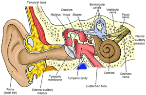
Three main compartments subdivide the human ear: the outer, middle and inner ear.
Outer Ear
The outer ear consists of the fleshy outer portion most commonly thought of when picturing the ear. This structure is referred to as the auricle or the pinna and is supported by cartilage. The auricle functions to funnel sound from the environment into the next section of the outer ear, the external auditory meatus. The external auditory meatus is the ear canal that leads to the tympanic window. The external auditory meatus is the passageway through the temporal bone and is coated in cerumen (earwax). The outer ear is exposed to the environment and is covered in skin. Earwax is produced in the outer ear in order to clean and lubricate the skin of the outer ear. Earwax contains a mixture of hydrocarbons, fatty acids, and cholesterol as well as a mix of antimicrobial proteins (Schwaab, 2011; Stransky, 2011). Earwax is produced by a combination of sebaceous and apocrine glands in the outer third portion of the outer ear (Alvord, 1997). The skin of the ear canal grows from inside to out and pushes skin cells to the exterior of the ear where it is eventually shed. This process expels the cerumen from the ear canal (Alberti, 1988). Human cerumen is dimorphic with wet and dry morphotypes (Matsunaga, 1962). The external auditory meatus terminates at the tympanic membrane (tympanic window or eardrum). The tympanic membrane is the thin membrane that separates the outer and middle ear (Alberti, 1988; Alvord, 1997).
Middle Ear
The middle ear, or tympanic cavity, is an air-filled cavity contain a set of three ossicles: the malleus, incus and stapes. The ossicles are conjoined sequentially with the malleus anchored to the tympanic membrane and the stapes anchored to the inner ear. The hammer, anvil, and stirrup respectively conduct the oscillations of the tympanic membrane from sound vibrations entering the outer ear to the inner ear via the oval window. The Eustachian tube connects the middle ear to the nasopharynx and functions to equilibrate air pressure between the middle and outer ear to prevent perforation of the ear drum. This is the reason that people can pop their ears by closing their mouth, plugging their nose and exhaling. The increase in pressure in the nasopharynx is transmitted into the middle ear via the Eustachian tube, causing the tympanic membrane to pop (Alvord, 1997; Alberti, 1988).
Inner Ear
The inner ear contains the organs and nerves that are involved in hearing and balance. The cochlea separates the inner and middle ear and is the snail-shaped auditory organ. The oval window of the cochlea vibrates as sound is conducted into the inner ear and the vibrations of the oval window. The perilymph inside the cochlea conducts the sound waves to the vestibular membrane. Inside of the vestibular membrane is endolymph fluid that conducts sound to the basilar membrane. Inside the basilar membrane, specialized hairs detect the sound waves and the action potentials created are sent to the brain via the vestibulocochlear nerves. The vestibule and semicircular canals function to maintain balance. The vestibule and semicircular canals sense the motion of the endolymph with specialized hair cells and assess the bodies position with respect to gravity. The action potentials are sent to the brain via the vestibulocochlear nerve. The endolymph and perilymph differ based on the potassium and sodium concentration. The endolymph contains higher concentration of potassium ions than sodium ions (Konishi, 1978). The difference in ion concentrations between the two presents a different environment to potential bacteria.
Microbiota of the Ear
Infections of the Ear
Include some current research, with at least one figure showing data.

Include some current research, with at least one figure showing data.

Include some current research, with at least one figure showing data.
References
[1] Hodgkin, J. and Partridge, F.A. "Caenorhabditis elegans meets microsporidia: the nematode killers from Paris." 2008. PLoS Biology 6:2634-2637.
Authored for BIOL 238 Microbiology, taught by Joan Slonczewski, 2015, Kenyon College.
