Magnetococcus marinus MC-1
Classification
Bacteria (Domain); Proteobacteria (Phylum); Alphaproteobacteria (Class); Magnetococcales (Order); Magnetococcaceae (family)
Species
Magnetococcus marinus MC-1
Description and Significance
Magnetotactic bacteria are morphologically, metabolically, and phylogenetically varying, but all have the ability to produce magnetosomes, which are biomineralized membrane-encased crystals of magnetite or greigite (Ji et al., 2017). MC-1 is a strain of Magnetococcus marinus, a magnetotactic coccus that is often found at the oxic-anoxic interface of bodies of water. MC-1 was first isolated from the oxic-anoxic interface of the Pettaquamscutt Estuary in Rhode Island, USA (Bazylinski et al., 2013).
Magnetotactic cocci are ubiquitous in freshwater, marine water, and mud samples from natural habitats. They are the most commonly encountered magnetotactic bacteria. Magnetococcus marinus are Gram negative, coccoid to ovoid in shape, and 1-2 µm in diameter.They are bilophotrichous, having two bundles of flagella on one side of the cell. Each cell contains magnetosomes, typically arranged in a linear chain inside the cytoplasm (Bazylinski et al., 2013).
As well as being ubiquitous in most water environments and being the most commonly encountered magnetotactic bacteria, Magnetococcus marinus has been tested in clinical studies for its potential use in drug delivery within tumors.
Genome Structure
Magnetococcus marinus MC-1 has a circular chromosome composed of 4719581 nucleotides, 3716 protein genes, and 57 RNA genes. The entire genome has been sequenced and is available from GenBank (Kegg Genome). The production of magnetosomes is a genetic process, the essential genes for which are clustered in a well-defined region called the Magnetosome Island (MAI) (Ji et al., 2017).
Magnetococcus marinus MC-1 and the magneto-ovoid strain, MO-1, are closely related, with a genetic similarity of 93.4% (Bazylinski et al., 2013). Both of these strains contain an extremely high amount of copies of flagellin protein-encoding genes (Zhang et al, 2012). Both the MC-1 and MO-1 strain have high proportions of genes associated with Alphaproteobacteria, Betaproteobacteria, Deltaproteobacteria, and Gammaproteobacteria, suggesting they are not closely related to any known proteobacteria and may represent a novel lineage, named Etaproteobacteria (Ji et al., 2017). However, both strains are still considered to be part of Alphaproteobacteria.
Cell Structure, Metabolism and Life Cycle
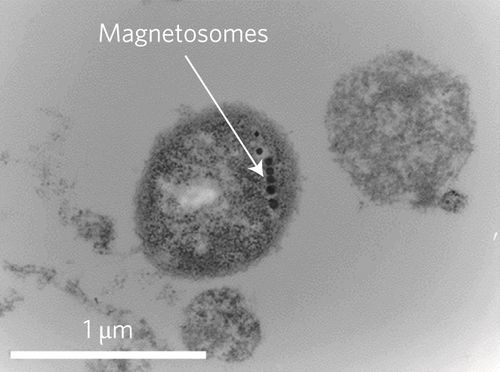
The cells of this strain of magnetotactic bacteria are 1-2 µm in diameter and are coccoid-ovoid shaped. Within the cell, there is a flagellar apparatus that consists of seven flagella surrounded by a sheath. They also contain microfibrils that are used to reduce friction to increase the speed of the cell. The microorganisms efficiently use the flagella and microfibrils to swim. Moreover, the cells contain magnetite crystals, called magnetosomes, which are enclosed in a membrane and are aligned in a chain. These crystals are necessary to align the bacteria with the earth’s magnetic field and are used by the microbe as a compass (Bazylinski et al., 2013). The chains of magnetosomes are also used to direct the bacteria to low levels of oxygen (Felfoul et al., 2016).
Cell structure of M. marinus showing internal magnetosome structures, taken using TEM (Felfoul).
M. marinus can grow either heterotrophically or autotrophically. Heterotrophically, the cultures utilize acetate as their electron donor. Autotrophically, the cultures utilize sulfide as the electron donor. This bacteria is capable of fixing nitrogen under both heterotrophic and autotrophic conditions (Bazylinski et al., 2013). This nitrogen fixation is important in the marine ecosystem, as it serves as a source of nutrients for other organisms.
This microorganism reproduces by the splitting of cells. During this splitting, the chain of magnetite crystals is split in half (Bazylinski et al., 2013).
Ecology and Pathogenesis
M. marinus is predominantly found in water containing vertical chemical stratification with a temperature range of 5-35 degrees Celsius. These habitats include: freshwater, marine water, and mud (Yan et al., 2012).This is an aerobic bacteria with the potential to survive (but not grow) in anaerobic environments (Bazylinski et al., 2013).
M. marinus is not known to cause any diseases in humans. However, they are used to help cancer patients. This type of bacteria deliver drug containing nanoliposomes to hypoxic zones in tumors. Hypoxic regions of cancerous tumors have been resistant to normal cancer treatments. The MC-1 strain is injected into patients and then is magnetically directed towards the tumor (Felfoul et al., 2016).
References
Bazylinski, Dennis A. et al. Magnetococcus marinus gen. nov., sp. nov., a marine, magnetotactic bacterium that represents a novel lineage (Magnetococcaceae fam. nov., Magnetococcales ord. nov.) at the base of the Alphaproteobacteria. International Journal of Systematic and Evolutionary Microbiology. 2013. Volume 63. p. 801–808.
Felfoul, Ouajdi et al. Magneto-aerotactic bacteria deliver drug containing nanoliposomes to tumor hypoxic regions. Nature Nanotechnology. 2016. Volume 11. p. 941-947.
Ji, Boyang et al. The chimeric nature of the genome of marine magnetotactic coccoid-ovoid bacteria defines a novel group of Proteobacteria. Environmental microbiology. 2017. Volume 19. p.1103-1119.
Magnetococcus marinus. (n.d.). Kegg Genome. Retrieved April 21, 2017, from http://www.genome.jp/dbget-bin/www_bget?gn%3AT00427
Yan, Lei et al. Magnetotactic bacteria, magnetosomes and their application. Microbiological Research. 2012. Volume 167. p. 507–519.
Zhang, S.D., Petersen, N., Zhang, W.J., Cargou, S., Ruan, J., Murat, D., et al. Swimming behaviour and magnetotaxis function of the marine bacterium strain MO-1. Environmental Microbiology. 2013. Volume 6. p. 14–20.
Author
Page authored by Melissa Mulford and Mariya Trenkinshu, students of Prof. Jay Lennon at IndianaUniversity.

