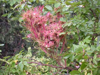
Rose Rosette Virus

Introduction
By Jacob Scharfetter
Rose rosette virus (RRV), also known as Rose rosette disease (RRD), is a viral plant pathogen. The symptoms of Rose Rosette Virus (RRV) was first recognized and recorded in Canada 77 years and has since become one of the most destructive diseases of commercial roses[1][2]. The plant pathogen RRV has only been found to affect the genus Rosa[3]. Most Rosa spp. are susceptible to RRV, making RRV a significant problem for landscapers and horticulturalists[4]. However, non-commercial, wild rose species of the Rosa genus, such as the meadow rose (R. blanda), swamp rose (R. palustris), Carolina rose (R. Carolina), prickly wild rose (R. acicularis), and burnet rose (R. spinosissima), show only minimal signs of susceptibility to RRV[4]. Currently, RRV is primarily distributed throughout the eastern United States ranging from the Eastern coasts of New England to the base of the Rocky Mountains[5]. In short, RRV is a destructive and highly lethal rose pathogen that expresses a significant threat to the commercial rose industry. This report seeks to highlight what we currently know about RRV and to highlight the areas where future research needs to be conducted.
Isolation History of RRV
The first indication that RRV was indeed a virus came when large virus-like particles were observed with scanning electron microscopy in Rosa multiflora and commercial roses in Northern Arkansas (Gergerich and Kim, 1983). In the same study, the double-membrane characteristic of the spherical envelope was observed for the large virus-like particles. The next breakthrough in the isolation of RRV came with the isolation of dsRNA from infected rose tissue (Di et al., 1990). dsRNA, being something that is prevalent and unique to viruses, strongly suggested that the causative agent for the rose rosette disease was a virus.
Up until 1995, rose rosette disease (RRD) was thought to be caused by a virus or a phytoplasma; a phytoplasma can be equally as small as a virus (Epstein and Hill, 1995). A phytoplasma was ruled out as the cause of rose rosette disease, by the lack of a DAPI DNA stain in isolated cells, no reversion in symptoms when plants were treated with tetracycline, and no amplification was detected using known primers of phytoplasmas via PCR analysis (Epstein and Hill, 1995).
The negative-sense RNA nature of RRV was finally elucidated in 2011, by using degenerate oligonucleotide primed reverse transcriptase PCR to amplify dsRNA (Laney et al., 2011). The purification of emaraviruses from inflected plants has been challenging to researchers due to the enveloped nature of the virus particles as well as by the low titre (Chulang Yu et al., 2013). This potentially explains why RRV and related emeraviruses were reported only having four genomic RNA segments rather than more (Mielke & Muehlbach 2007; Elbeaino et al., 2009; Laney et al., 2011). Recently, from more sensitive analysis another three RNA segments were isolated and detected in RRV (Di Bello et al., 2015). There are many things not fully understood about RRV. At the foremost of this list is the pathogenicity of RRV. Future studies need to be conducted in order to elucidate the mechanism of entry for RRV, the replication of RRV, and the possible latency of RRV.
Virion and Genome Structure
The rose rosette viron particle is of large size ranging from 120-150nm (Gergerich and Kim, 1983). The RRV viron particle is comprised of symmetrically helical enveloped ribonucleocapsid and has been described as having a spherical shape (Epstein and Hill, 1994; Kim et al., 1995). Rose rosette virus is a negative-sense RNA virus and was identified in 2011 as a member of the genus, Emaravirus (Laney et al., 2011). Like European mountain ash ringspot associated virus (EMARaV), RRV has four common RNA coding segments, RNA1-RNA4 (Ishikawa et al., 2013) as well as three other uncharacterized RNA5-7 segments (Di Bello et al., 2015). The RNA1-4 segment protein products are referred to p1-p4 respectively (see Figure 4; Laney et al. 2011). In RRV the RNA1, RNA2, and RNA3 each contain an open reading frame (OPR) that putatively encodes for RNA-dependent RNA polymerase (RdRp, RNA1), glycoprotein precursor (RNA2), and nucleocapsid (RNA3) (Mielke-Ehret & Muehlbach 2012) . Fascinatingly, segmental RNA from the RRV genome was found to be uncapped, but mRNA of RRV transcripts were found to be capped with 7-methylguanylate just like all eukaryotic mRNA transcripts (Laney et al., 2011).
In RRV, RNA4 (p4) function has not be elucidated. However, RRV p4 is closely related to virus raspberry leaf blotch emaravirus p4 (RLBV, see Figure 2) (McGavin et al., 2012). In a study looking at p4 in RBLV, it was shown that the p4 protein localizes to the plasmodesmata, hinting that the protein is a viral movement protein otherly known as an MP (Chulang Yu et al., 2013). Upon further investigation the p4 protein from RLBV rescued cell-to-cell movement of a MP-deficient potato virus X (PVX), which provides evidence that the p4 protein in RRV is likely an MP protein (Chulang Yu et al., 2013). There are direct genetic indications that RRV p4 is a cell-to-cell movement protein with the largest piece of evidence coming from the fact that there are dnaK and ATPase motifs in the RRV RNA4 segment, which codes for p4 (Laney et al., 2011). dnaK and ATPase domains are required for plant viral movement proteins (Alzhanova et al., 2001). It is likely that p4 is a movement protein. However, until an experiment like that Chulang Yu and colleagues (2013) is conducted in an RRV infected host, we will not know for certain the function of RRV p4.
Unlike RRV, other emaraviruses such as RLBV has at least eight putatively encoding RNA segments (McGavin et al., 2012). Due to the low titre and enveloped nature of RRV, RRV may be comprised of more RNA segments (Laney et al., 2011; Chulang Yu et al., 2013). In a follow-up isolation study by Di Bello and colleagues (2015), three new RNA genome segments were found. However, it is possible fragile or low concentration RNA segment regions may have gone undetected in RRV samples. In order to build a better map of the RRV genome, future dsRNA isolation studies of RRV will have to be conducted in order to confirm that there are only seven RNA segments.
Section 3
Include some current research, with at least one figure showing data.
Section 4
Conclusion
References
- ↑ Conners, I.L. Twentieth Annual Report of the Canadian Plant Disease Survey 1940; Department of Agriculture: Ottawa, Canada, 1941; p. 98.
- ↑ Laney A., Keller K., Martin R.,& Tzanetakis I. A discovery 70 years in the making: characterization of the Rose rosette virus. 01 July 2011, Journal of General Virology 92: 1727-, doi: 10.1099/vir.0.031146-0
- ↑ Dobhal, S., Olson, J. D., Arif, M., Suarez, J. A. G., & Ochoa-Corona, F. M. (2016). A simplified strategy for sensitive detection of Rose rosette virus compatible with three RT-PCR chemistries. Journal of virological methods, 232, 47-56. doi:10.1016/j.jviromet.2016.01.013
- ↑ 4.0 4.1 Epstein, A.H., & Hill, J.H. (1999). Status of rose rosette disease as a biological control for multiflora rose. Plant disease, 83(2), 92-101. doi:10.1094/PDIS.1999.83.2.92
- ↑ Windham, M., Windham, A., Hale, F., & Amrine Jr, J. (2014). Observations on Rose rosette disease.
Authored for BIOL 238 Microbiology, taught by Joan Slonczewski, 2017, Kenyon College.
