File:Image0 (2).jpeg
From MicrobeWiki, the student-edited microbiology resource

Size of this preview: 479 × 599 pixels. Other resolution: 700 × 876 pixels.
Original file (700 × 876 pixels, file size: 241 KB, MIME type: image/jpeg)
Summary
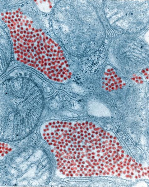
This 83,900X magnified image shows a salivary gland tissue section extracted from a eastern equine encephalitis (EEE) virus infected mosquito. This image was taken using transmission electron microscopy, and the EEE virus particles are digitally colorized in red. Photo by Fred Murphy and Sylvia Whitfield. (21)
File history
Click on a date/time to view the file as it appeared at that time.
| Date/Time | Thumbnail | Dimensions | User | Comment | |
|---|---|---|---|---|---|
| current | 15:17, 7 December 2020 |  | 700 × 876 (241 KB) | Ocnieves (talk | contribs) | This 83,900X magnified image shows a salivary gland tissue section extracted from a eastern equine encephalitis (EEE) virus infected mosquito. This image was taken using transmission electron microscopy, and the EEE virus particles are digitally colori... |
You cannot overwrite this file.
File usage
The following page uses this file:
