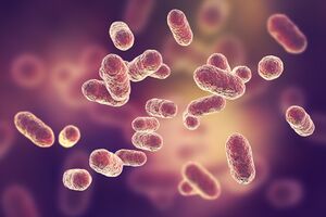
Porphyromonas Gingivalis and Gum Disease
Introduction

By Babiker Higazi
The abbreviation PG might bring to mind a family-friendly movie for the average person, but for periodontists, it evokes a different sentiment. This is because Porphyromonas gingivalis, commonly known as PG, is a leading cause of gum disease worldwide [1]. Gum disease or periodontal disease is often the result of infections in the gums and bones that support teeth [1]. Along with tooth decay, it poses one of the most significant threats to dental health. According to a recent report, around 47% of adults over the age of 30 have some form of periodontal disease, which is more commonly found in men and people living below the federal poverty line [1]. P. gingivalis is an anaerobic, gram-negative bacterium and an integral bacteria in the oral microbiota. Along with many periodontal conditions, P. gingivalis has even been linked to Alzheimer's disease in the world of neuroscience [2]. To better understand this bacterium, let's trace its journey from biofilm formation to the onset of devastating diseases.".
The insertion code consists of:
Double brackets: [[
Filename: PHIL 22882 lores.jpg
Thumbnail status: |thumb|
Pixel size: |300px|
Placement on page: |right|
Legend/credit: Electron micrograph of the Ebola Zaire virus. This was the first photo ever taken of the virus, on 10/13/1976. By Dr. F.A. Murphy, now at U.C. Davis, then at the CDC. Every image requires a link to the source.
Closed double brackets: ]]
Other examples:
Bold
Italic
Subscript: H2O
Superscript: Fe3+
Sample citations: [1]
[2]
A citation code consists of a hyperlinked reference within "ref" begin and end codes.
To repeat the citation for other statements, the reference needs to have a names: "<ref name=aa>"
The repeated citation works like this, with a forward slash.[1]
Biofilm Formation and Intracellular Invasion
P. gingivalis, like many bacteria in the oral cavity, relies on the formation of biofilm for its virulence. The oral cavity is composed of a wide range of substrates, including soft tissue, enamel, and even implanted materials. P. gingivalis has developed an intricate mechanism for biofilm formation, allowing it to adhere to a diverse range of materials. The bacterium expresses two types of fimbriae, which are proteinaceous appendages anchored to the outer membrane of bacteria [3]. One of these fimbriae is long and composed of FimA protein subunits [3]. These fimbriae assist P. gingivalis in its initial attachment to host cells [4]. After associating with the host cell, P. gingivalis coats the surrounding area with capsular polysaccharides [5]. The coating creates a barrier between P. gingivalis and other surface components, such as proteins and lipopolysaccharides [4].
P. gingivalis biofilms tend to be mixed, meaning they also contain other bacterial species. Thus, P. gingivalis has adapted to use quorum sensing as a tool for biofilm formation [5]. Specifically, the LuxS/Autoinducer-2 system allows P. gingivalis to communicate with neighboring species [6]. This communication is essential, as it allows P. gingivalis to know when it has reached its critical mass, along with neighboring species. At this point, the mixed bacteria coordinate their extracellular matrix formation, dramatically increasing the likelihood of successful biofilm formation. P. gingivalis interactions with neighbors are also beneficial from a nutritional standpoint. Another major oral bacterium, T. denticola, often occupies the same areas as P. gingivalis. P. gingivalis secretes isobutyric acid, which enhances the growth of T. denticola [7]. Meanwhile, T. denticola produces succinate, which enhances the growth of P. gingivalis [7]. These interactions may explain why P. gingivalis and T. denticola show enhanced biofilm formation when cultured together [8].
The biofilm mechanisms enable P. gingivalis to thrive in subgingival dental plaque. However, this bacterium can also be found in other periodontal sites, such as the epithelial gingival cells of our periodontal tissue [9]. Gingival cells are an important component of our innate immune system, acting as a barrier to prevent bacterial invasion of the periodontium [10]. Despite this, P. gingivalis has been found to penetrate this barrier and invade the deeper tissue layers of the periodontium [9]. In these layers, P. gingivalis faces less pressure from immune defenses, allowing it to proliferate, multiply, and spread into adjacent tissues [11].
The intracellular invasion process of P. gingivalis consists of four steps: adhesion, entry, intracellular trafficking, and exit [12]. First, P. gingivalis uses its FimA fimbriae to attach to the alpha-5-beta-1 integrin on the surface of gingival cells [13]. It then exploits the cellular endocytosis pathways used by other molecules to enter the cell [9]. Once inside the host cell, P. gingivalis disrupts essential cellular functions such as proliferation and cellular migration. After causing severe damage to the function of the gingival cells, P. gingivalis may exit the cell via various exocytotic pathways. Some of these pathways lead to the bacterium being sorted into lytic compartments, such as lysosomes and autolysosomes [14]. Others allow P. gingivalis to enter a recycling pathway, where functional proteins and lipids from the bacteria are utilized by the gingival cells [14].
Every point of information REQUIRES CITATION using the citation tool shown above.
Section 2
Include some current research, with at least one figure showing data.
Section 3
Include some current research, with at least one figure showing data.
Section 4
Conclusion
References
Authored for BIOL 238 Microbiology, taught by Joan Slonczewski, 2023, Kenyon College
