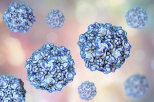
Feline panleukopenia virus
Classification
Viruses; Monodnaviria; Shotokuvirae; Cossaviricota; Quintoviricetes; Piccovirales; Parvoviridae; Parvovirinae; Protoparvovirus; Protoparvovirus carnivoran 1; Feline panleukopenia virus (genogroup)
Species
|
NCBI: [1] |
Protoparvovirus Protoparvovirus carnivoran 1
Description and Significance
Feline panleukopenia virus (FPV or FPLV) is a non-enveloped, icosahedral, single-stranded DNA virus and is sometimes referred to as feline distemper, cat plague, or feline parvovirus. FPV was first identified in the 1920s and has maintained genomic stability to a degree. FPV is a serious, highly contagious and potentially fatal issue among unvaccinated felines. Following proper vaccine guidelines can prevent felines from getting infected.
Genome Structure
The genome of feline panleukopenia virus contains approximately 5123 nucleotides and 4 open-reading frames that encode NS1 and NS1 (non-structural proteins) and VP1 and VP2 (structural proteins). The first open-reading frame contributes to replication, transcription and cell death of the host's cells. The second open-reading frame directs the formation and translation of capsid proteins. The third open-reading frame consist of VP2's sequence and contributes to cell infection and nuclear transport of capsids. The fourth open-reading frame encodes VP2 which is considered major capsid protein since it makes up approximately 90% of the whole capsid. The amount of base pairs in the DNA sequence varies depending on the specific strain of the virus. Due to the Y-type structure at the 3-terminal and the U-shaped structure at the 5-terminal, it is challenging for researchers to amplify the full-length genome of this virus and many others in the parvovirus genus.
Cell Structure, Metabolism and Life Cycle
Parvoviruses, including FPV, do not have DNA polymerases and cannot promote cell division. FPV relies on its host for its metabolic processes and use the host's cell division machinery, specifically cells in S-phase, to complete their lytic cycle. The virus usually attacks bone marrow, lymphatic tissue, and intestinal epithelial cells. The virus has an incubation period of 2 to 7 days. It can remain in an environment for days to months and possibly years. Due to its resistance to most disinfectants it is a difficult virus to eliminate from the environment making it more possible for other vulnerable animals to contract it.
Ecology and Pathogenesis
Habitat; symbiosis; biogeochemical significance; contributions to environment.
If relevant, how does this organism cause disease? Human, animal, plant hosts? Virulence factors, as well as patient symptoms.
The virus can be transmitted either by the fecal-oral route or by indirect transmission via fomites contaminated by infected individuals, as well as from humans that have the virus on their hands or clothes. The virus can live in an environment for months to a year and in some instances over a year. FPV is also highly resistant to certain disinfectants making it difficult for owners to remove it from their household. Using transferrin receptors FPV can enter cells and replicate inside of cells that are currently in the synthesis phase of mitosis. FPV can occur in cats younger than 1 year old, or unvaccinated or improperly vaccinated cats of any age. Severity of the virus depends on age, immunity of the individual and infections from other bacteria or viruses. FPV causes feline infections enteritis (FIE) which is a severe infection of the gut that is fatal and highly contagious making it spread rapidly in a multiple cat household. In addition to domestic cats, FPV can infect wild cats, raccoons, foxes, and mink. Clinical symptoms of infected individuals are lethargy, diarrhea, inappetence, fever (103°F to 107°F or 39.5°C to 42.5°C), vomiting, weight loss. In severe cases, cats can have ulcers in the mouth as well as exhibit mucosal pallor. In terminal cases, cats can be hypothermic, bradycardic, and comatose. If left untreated or if the virus has progressed too far then death can occur usually because of dehydration, low blood sugar, imbalanced electrolytes, hemorrhaging, or bacteria in the bloodstream.
References
Author
Page authored by Phoebe Chambers, student of Prof. Bradley Tolar at UNC Wilmington.

