Equine Development of Gut Microbiota
In utero, horses intestinal tracts are close to sterile. Soon after birth, however, microbial colonization skyrockets. Proper colonization lasts about 50 days, and is incredibly important as improper microbial gut content can result in inflammation and metabolic disease [1]. Equine hind-gut microbiota enable nutritional optimization from an otherwise nutrient-poor foraging diet via plant material fermentation [2]. The initial colonization, stabilization, and then weaning period (4-6 months old) as a foal transfers to solid food are important periods in establishing the microbial composition of the colon [1].

By Mira Allen
Topic: equine development of gut microbiota
The insertion code consists of:
Double brackets: [[
Filename: PHIL_1181_lores.jpg
Thumbnail status: |thumb|
Pixel size: |300px|
Placement on page: |right|
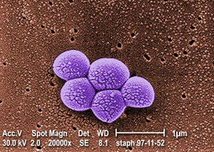
Legend/credit: Magnified 20,000X, this colorized scanning electron micrograph (SEM) depicts a grouping of methicillin resistant Staphylococcus aureus (MRSA) bacteria. Photo credit: CDC. Every image requires a link to the source.
Closed double brackets: ]]
Other examples:
Bold
Italic
Subscript: H2O
Superscript: Fe3+
Sample citations: [1]
[2]
A citation code consists of a hyperlinked reference within "ref" begin and end codes.
To repeat the citation for other statements, the reference needs to have a names: "<ref name=aa>"
The repeated citation works like this, with a forward slash.[1]
Section 1
Include some current research, with at least one figure showing data.
Every point of information REQUIRES CITATION using the citation tool shown above.
Section 2
Include some current research, with at least one figure showing data.
Section 3
Include some current research, with at least one figure showing data.
Section 4
Conclusion
References
- ↑ 1.0 1.1 1: https://doi.org/10.1038%2Fs41598-019-50563-9
2: https://doi.org/10.1186/s42523-019-0013-3
Hodgkin, J. and Partridge, F.A. "Caenorhabditis elegans meets microsporidia: the nematode killers from Paris." 2008. PLoS Biology 6:2634-2637.] - ↑ Bartlett et al.: Oncolytic viruses as therapeutic cancer vaccines. Molecular Cancer 2013 12:103.
Authored for BIOL 238 Microbiology, taught by Joan Slonczewski,at Kenyon College,2024
