Rickettsia rickettsii
A Microbial Biorealm page on the genus Rickettsia rickettsii
Classification
Higher order taxa
Bacteria; Proteobacteria; Alpha Proteobacteria; Rickettsiales; Rickettsiaceae; Spotted fever group
Species
|
NCBI: Taxonomy |
Rickettsia rickettsii
Other Names
synonym: Dermacentroxenus rickettsii
synonym: "Dermacentroxenus rickettsii" Wolbach 1919
synonym: Rickettsia rickettsii (Wolbach 1919) Brumpt 1922
Description and significance

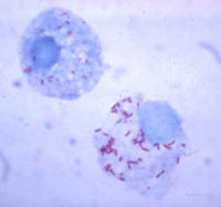
Rickettsia rickettsii is a small, rod-shaped bacterium known to cause Rocky Mountain spotted fever (RMSF). This disease can be transmitted to humans either from a tick bite with an incubation period of 1 week, or by contamination of a cut on the skin or a wound with ticks feces. Dr. Ricketts first isolated this microbe in Montana, 1906. Rickettsia need host cells to be able to grow. In addition, they also require arthropods as vectors; therefore, ticks are the vectors used for Rickettsia rickettsii. [2] [4] [6]
Genome structure
The unfinished genome sequencing of Rickettsia rickettsii is made up of 1,257,710 nucleotides long. There are 1311 genes, and all of them code for proteins. The guanine and cytosine content is 32% of its DNA. Rickettsia rickettsii has no pseudo genes, and it’s made up of DNA molecules. The topology of Rickettsia rickettsii is neither circular or linear chromosomes. In addition, no known plasmids have been found. [3]
Infection with Rickettsia rickettsii also triggers an active response that alters cellular phenotypes. There is an increase in the secretion of cytokines and proteins. During Rickettsia rickettsii infection, nuclear factor-kappa B (NF-KB) is activated. This is a group of transcription factors that regulate cell adhesion, proliferation, the immune system, inflammation, stress responses, and host pathogenic interactions. [10]
Cell structure and metabolism

The Rickettsia rickettsii bacterium are intracellular organisms, and they live in the cytoplasms of host cells sometimes in the nucleus. They are adapted as intracellular organisms because they have transport systems and metabolic enzymes. These microbes divide by binary fission and take about 8 hours to double. [2]
Rickettsia rickettsii are similar to small, gram-negative nods. They are barely visible under a light microscope, and they stain poorly with Gram stains. The cell wall is made up of peptidoglycan, lipopolysaccharide, and a cell membrane. These microbes contain two outer membrane proteins, outer membrane protein A (OmpA) and outer membrane protein B (OmpB). OmpA sticks to the host cell wall, and it is a protective layer of about 190 kDa. It contains a region of 13 identical repeating units that include antigenic function. OmpB is an abundant outer membrane protein that contains sequences related to the typhus group of rickettsiae.[2][7]
Ecology
Rickettsia rickettsii can be found in the western hemisphere, more notably in North America and South America. In North America, it’s more well known in the southeast and southcentral states. In North America, Rickettsia rickettsii is transmitted by the American dog tick (Dermacentor variabilis) and the Rocky Mountain wood tick (Dermacentor andersoni). In Latin America. In Latin America, Rickettsia rickettsii is transmitted by Rhipicephalus sanguineus and the Cayenne tick Amblyomma cayenne. [9]
Describe any interactions with other organisms (included eukaryotes), contributions to the environment, effect on environment, etc.
Pathology
Typical symptoms of RMSF can appear 2 to 14 days after exposure and include fever, headache, depression, nausea, vomiting, and gradually may develop a skin rash called purpura or petechiae. Sometimes the rash occurs 2 to 5 days after the onset of the fever. Serious cases of RMSF can include central nervous system, pulmonary, or hepatic injuries. Patients that have compromised immune systems often have an increased susceptibility to other infections. [2][9]
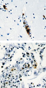
Rickettsia Rickettsii can infect endothelial cells within the human body through the vascular smooth muscle cells. [5] In addition, it is an intracellular pathogen that can infect and multiple within the nucleus or cytosol of endothelial cells of the blood vessels. [9]
How does this organism cause disease? Human, animal, plant hosts? Virulence factors, as well as patient symptoms.
Application to Biotechnology
Does this organism produce any useful compounds or enzymes? What are they and how are they used?
Current Research
Currently there is a major lack in controlling the RMSF organism. In addition, an effective diagnostic test to detect early symptoms is still not available. Doctors have a hard time detecting RMSF because the patients rarely have the Rickettsia rickettsii antibodies. Although PCR (Polymerase Chain Reaction) testing is available in a few laboratories, detection of Rickettsia DNA in the blood is still tactless, especially in the early symptoms. After diagnosis with RMSF, it is usually treated with Tetracycline unless the CNS has already been affected thereby using Chloramphenicol instead.[8] [9]
Enter summaries of the most recent research here--at least three required
References
1) NCBI Taxonomy Browser "Rickettsia Rickettsii" Retrieved 26 August, 2007
2) Primary Care Update for OB/GYNS, Volume 9, Issue 1, January-February 2002, Pages 28-32 Whitney Jamie
3)http://www.ncbi.nlm.nih.gov/sites/entrez?Db=genome&Cmd=ShowDetailView&TermToSearch=5211
4) http://jmm.sgmjournals.org/cgi/content/full/56/1/138
6) Phylogenetic analysis of the genus Rickettsia by 16S rDNA sequencing Research in Microbiology, Volume 146, Issue 5, June 1995, Pages 385-396 V. Roux and D. Raoult
7) Comparison of the rompA gene repeat regions of Rickettsiae reveals species-specific arrangements of individual repeating units Gene, Volume 125, Issue 1, 15 March 1993, Pages 97-102 R. D. Gilmore
8) http://www.cdc.gov/ncidod/eid/vol12no04/05-1282.htm
9) Transfusion-transmitted tick-borne infections: A cornucopia of threats Transfusion Medicine Reviews, Volume 18, Issue 4, October 2004, Pages 293-306 David A. Leiby and Jennifer E. Gill
Edited by Amber Chou; student of Rachel Larsen
