Synechococcus
Classification
Higher order taxa:
Bacteria; Cyanobacteria; Chroococcales; Synechococcus
Species:
Synechococcus elongatus, Synechococcus WH 8102
Description and Significance
Synechococcus sp. are cyanobacteria, oxygenic phototrophs that can photolyze either H2O or H2S. Synechococcus is the main source of primary production in oligotrophic, pelagic marine waters. Their can cause destructive blooms, producing neurotoxins. Their growth is generally limited however by the concentration of nutrients and trace metals such as iron and phosphorus.
Cyanobacteria: General Information
Cyanobacteria as a whole are distinguished form other eubacteria by their ability to carry out oxygenic photosynthesis, closely resembling that of eukaryotic photoautotrophs. Like eukaryotic microalgae, they have two connected photosystems that provide energy for carbon fixation in the Calvin cycle. Cyanobacteria, however, have different light harvesting complexes. In eukaryotes, in addition to the chlorophyll a found in photosystems I and II, they have other accessory chlorophylls in their light harvesting complexes. Cyanobacteria have phycobilins, instead of these accessory chlorophylls, which absorb light over a much wider range of wavelengths. Cyanobacteria, although viewed unkindly for their destructive blooms (often producing neurotoxins) and oppressive stench, are essential components of even oligotrophic systems. Many genera of cyanobacteria thrive in nutrient-rich, eutrophic environments, and are, as such considered indicators of eutrophication, which currently troubles many aquatic ecosystems.
Genome Structure
Currently there are two complete sequencing of Synechococcus genome. One, Synechococcus sp. WH 8102, has 2434428 bp and has one chromosome. The other, Synechococcus elongatus PCC 6301, has 2696255 bp and has one chromosome.There are also 9 other genome projects that are in progress/draft assembly.
Cell Structure and Metabolism

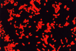
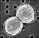
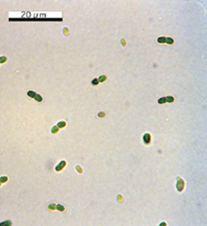
Synechococci are found either as solitary cells or in small clusters or pairs. They are spherical or ellipsoidal in shape and are photoautotrophic. They are characterized as euryphotic and are capable of growth at a wide range of light intensities. They prefer neutral to slightly alkaline pH and are motile organisms, yet have no flagella. In the absence of flagella, the cell surface must generate thrust, and does so by oscillating in the manner of a traveling wave. One distinct biochemical feature of Synechococcus is the presence of phycoerythrin, a pigment whose orange fluorescence can be detected at an excitation wavelength of 540 nm, and serves to identify Synechococcus.
Ecology

Synechococci are a significant form of primary production worldwide, although their contribution to primary production usually increases in oligotrophic environments, and they are essential in supporting upper trophic levels. Synechococcus colonies are found only in salt water, and contribute about 25% of primary production in pelagic waters. Although they are non bloom-forming they reach their highest abundance during periods of severe nutrient limitation in summer and autumn. They are usually limited by nitrogen as opposed to phosphorus. It is thought that their habit of clustering together to form tightly packed colonies aids in more efficient nutrient recycling, enabling them to prosper under depleted conditions. In both marine and freshwater strains of Synechococcus growth and division are light-dependent, and operate on what can be called a circadian rhythm. Cell division reaches a maximum in the afternoon generating an increase in cell number that proceeds into the night, when the number of cells gradually declines. These light/dark cycles create a rhythmic pattern of cell division and related growth, which is driven by prevailing light conditions. Optimum growth occurs under very low light conditions, but they also have a method of photo-adatation which permits cell growth and photosynthetic activity to continue at very high irradiances.
Phages
When phages infect cyanobacteria, they are called cyanophages. Syenchococcus is infected via lysogeny, when the viral genome is stably maintained as a prophage within its host. There is a seasonal pattern to the interaction between Synechococcus and its phage, which is consistent with the occurrence of lysogeny at times of low host availability, resource limitation, or adverse environmental conditions in order to ensure viral survival. Lysogeny most often occurs in synechococcus during the summer months, and may be due to an increase in ultra-violet light or temperature. When the viruses cause lysis as opposed to lysogeny, they can be important in keeping Synechococcus numbers under control.
References
Fahnenstiel, Gary L. & Hunter J. Carrick. 1991. Physiological characteristics and food-web dynamics of Synechococcus in Lakes Huron and Michigan. Limnology and Oceanography, Vol. 36: 219-234.
>Webb, E. Cyanobacteriology at WHOI. Woods Hole Oceanographic Institution.

