Rickettsia
A Microbial Biorealm page on the genus Rickettsia
Classification
Higher order taxa:
Bacteria; Proteobacteria; Alphaproteobacteria; Rickettsiales; Rickettsiaceae; Rickettsieae
Species:
Orientia tsutsugamushi;
spotted fever group: Candidatus R. principis; Israeli tick typhus rickettsia; R. aeschimannii; R. africae; R. akari; R. amblyommii; R. andeana; R. australis; R. conorii; R. cooleyi; R. felis; R. heilongjiangensis; R. heilongjiangii; R. helvtica; R. honei; R. hulinensis; R. hulinii; R. japonica; R. martinet; R. massiliae; R. monacensis; R. montanensis; R. moreli; R. parkeri; R. peacockii; R. rhipicephali; R. rickettsii; R. sibirica subgroup; R. slovaca; R. sp.
Typhus group: R. canadensis; R. prowazekii; R. typhi
Unclassified Rickettsia: Candidatus R. tarasevichiae; R. bellii; R. publicis; R. sp.
|
NCBI: Taxonomy Genome: -R. conorii str. Malish 7 -R. prowazekii str. Madrid E |
Description and Significance
Rickettsia bacteria are well known pathogens. Rickettsia conorii causes Mediterranean spotted fever in humans and is contracted by contact with infected brown dog ticks. Other Rickettsia include Rickettsia prowazekii, which causes typhus, R. rickettsii, which causes Rocky Mountain spotted fever, and Rickettsia akari, which causes rickettsialpox. In addition to this, the genome of Rickettsia prowazekii is similar to mitochondrial genomes; phylogenetically, R. prowazekii is more closely related to the mitochondria than any other microbe known thus far.
Genome Structure
The genome of Rickettsia prowazekii is 1,111,523 base pairs in length and contains 834 protein-coding genes. It contains no genes for anaerobic glycolysis as well as genes involved in the biosynthesis and regulation of biosynthesis of amino acids and nucleosides in free-living bacteria similar to mitochondrial genomes. Unlike the mitochondrial genome, however, the genome of R. prowazekii contains a complete set of genes encoding for the tricarboxylic acid cycle and the respiratory-chain complex. Still, the genomes of Rickettsia as well as the mitochondria are small, highly derived, "products of several types of reductive evolution" (Andersson et al. 1998).
Cell Structure and Metabolism
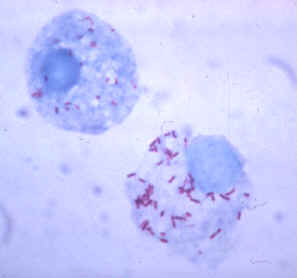
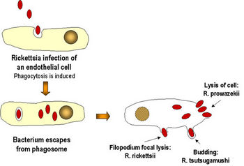
Rickettsia bacteria are obligate intracellular pathogens that are dependent on entry, growth, and replication within the cytoplasm of a eukaryotic host cell. The host cell then lysis and releases the rickettsial progeny to initiate a new infection cycle. The infection generally doesn't result in complete shutdown of the host machinery. Apparently, "vigorous host responses" generally clear the rickettsial pathogens (Radulovic et al. 2001). Conversely, the host's immune responses can also lead to the persistence of a subclinical infection even years past primary infection and/or antibiotic treatment. One theory on how rickettsiae survives in host cells has to do with the "suppression of the antimicrobial activities of the eukaryotic target cells, specifically monocytes/macrophages" (Radulovic et al. 2001).
Ecology
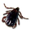
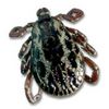
Male and female brown dog ticks (Rhipicephalus sanquineus) that are known to carry Rickettsia. From the Texas Department of Health |
Rickettsia bacteria are commonly carried by anthropods like ticks, mites, lice, or fleas. Another way for animals and humans to contract the bacteria is through wild rodents that have been infected with a Rickettsia bacteria by these louse. Different forms of Rickettsia and the diseases that they cause can be found all over the world. While some diseases themselves are worldwide, others, such as Oriental spotted fever caused by R. japonica in Japan, are localized to one general place or area.
Pathology
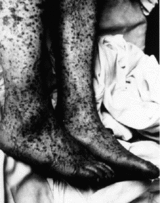
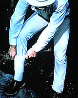
R. prowazekii is most known for being the agent of epidemic, louse-borne typhus in humans. It has infected approximately 20-30 million humans during World War I and killed another few million after World War II (Andersson et al. 1998). Typhus 'ranks as one of the main epidemic diseases of human history, a truly apocalyptic pestilence that follows in the wake of wars, famine, and other human misfortune" (Gray 1998). Rocky Mountain spotted fever, which is caused by infection with R. rickettsii, is the most severe rickettsial illness that is tickborne in the US. The primary ticks that carry it are the American dog tick (Dermacentor variabilis) and the Rocky Mountain wood tick (Dermacentor andersoni). Patients infected with R. rickettsii generally have nonspecific symptoms including fever, nausea, vomiting, muscle pain, lack of appetite, and severe headache after an incubation period about 5-10 days following an infected tick bite. Later symptoms include rash, abdominal pain, joint pain, and diarrhea. Fever, rash, and a previous tick bite are usually the most common components of clinical diagnosis. Rocky Mountain spotted fever is treated by a tetracycline antibiotic like doxycycline; once a person has had the disease, they are thought to have long lasting immunity against re-infection (CDC).
Below are tables of different groups of Ricketsia along with the diseases that each species cause and their general geological distribution. From The University of South Carolina.
Spotted Fever Group
| Organism | Disease | Distribution |
| R. rickettsii | Rocky Mountain spotted fever | Western hemisphere |
| R. akari | Rickettsialpox | USA, former Soviet Union |
| R. conorii | Boutonneuse fever | Mediterranean countries, Africa, India, Southwest Asia |
| R. sibirica | Siberian tick typhus | Siberia, Mongolia, nothern China |
| R. australis | Australian tick typhus | Australia |
| R. japonica | Oriental spotted fever | Japan |
Typhus Group
| Organism | Disease | Distribution |
| R. prowazekii |
Epidemic typhus |
South America and Africa Worldwide United States |
| R. typhi | Murine typhus | Worldwide |
Scrub typhus group
| Organism | Disease | Distribution |
| R. tsutsugamushi | Scrub typhus | Asia, northern Australia, Pacific Islands |
References
General:
- Reinert, Birgit. 2001. "Insights into genome evolution: The sequence of Rickettsia conorii." Genome News Network
- Andersson, Siv G. E., Alireza Zomorodipour, Jan O. Andersson, Thomas Sicheritz-Ponten, U. Cecilia M. Alsmark, Raf M. Podowski, A. Kristina Naslund, Ann-Sofie Eriksson, Herbert H. Winkler, and Charles G. Kurland. 1998. "The genome sequence of Rickettsia prowazekii and the origin of mitochondria." Nature, vol. 396. Macmillan Publishers Ltd. (133-143)
Cell Structure and Metabolism
- Radulovic S., R. W. Price, M. S. Beier, J. Gaywee, J. A. Macaluso, and A. Azad. 2002. "Rickettsia-macrophage interactions: Host cell responses to Rickettsia akari and Rickettsia typhi." Infection and Immunity, vol. 70, no. 5. American Society for Microbiology. (2576-2582)
Pathology:
- Dr. Gene Mayer. University of South Carolina: Rickettsia, Ehrlichia, Coxiella, and Bartonella
- CDC: Rocky Mountain spotted fever
- Gray, Michael W. 1998. "Rickettsia, typhus and the mitochondrial connection." Nature, vol. 396. Macmillian Publishers Ltd. (109-110)
