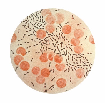
Streptococcus Pneumoniae in China
Classification
Higher Order Taxa
Bacteria (domain); Firmicutes (phylum); Bacilli (class); Lactobacillales (order); Streptococcaceae (family); Streptococcus (genus)
Species
Streptococcus pneumoniae
Also known as pneumococcus
Introduction and Significance
Streptococcus pneumoniae proves to be one of the most prevalent bacterial infections in humans. Discovered by Louis Pasteur in the early 1880s, S. pneumoniae was diagnosed as one of the main causes of pneumonia, along with meningitis and bacteremia. Traveling from the host to the uninfected primarily through respiratory droplets, S. pneumoniae infects both children and adults, being known notoriously as a cause of death for children in China. Two vaccines are currently used as preventative measures, while antibiotics are prescribed for existing infections. Unfortunately, overuse of antibiotics has led to universal resistance and will soon require new innovations in treatment.
S. pneumoniae was used in the revolutionary discovery of transformation, the uptake of DNA by a cell. In 1920s, while attempting to create a vaccine, Frederick Griffith used two strains of S. pneumonia bacteria, a smooth strain, with a polysaccharide capsule that caused pneumonia upon injection, and rough strain, that lacked this capsule and did not cause pneumonia when injected into mice. (20) The rough strain was observed to cause disease when injected with an attenuated smooth strain, indicating an acquirement of smooth trait by the rough strain. Oswald Avery, Colin MacLeod, and Maclyn McCarty demonstrated that the transforming factor in Griffith's experiment was DNA, not protein as was widely believed at the time. (3)
Genome Structure
The genome of Streptococcus pneumoniae TIGR4, a very recently sequenced strain, is 2,160,842 base pairs long. The circular double-stranded DNA has a guanine-cytosine content of 39%, and thus a 61% adenosine-thymine content. 83% of the genome is coding sequence corresponding to 2232 genes, 2106 of which code for protein. (19)
Genomic statistics vary between different strains, in accordance with varying virulence. Since the introduction of antibiotics, Streptococcus pneumoniae has displayed major increase in resistance to them. This is likely due to such gene transfer mechanisms as transformation and conjugation, as well as mutations that are selected for in the presence of antibiotics.
Cell Structure and Metabolism
Streptococcus pneumoniae is a non-motile, non-spore forming, lancet-shaped mesophile that grows in pairs or short chains. It is classified as Gram-positive, having a cell membrane surrounded by a thick cell wall. There are over 90 different antigenic serotypes, classified based on capsid antigens. (14) Three major surface layers encompass the cell, all of which play important roles in the bacterium’s success as a pathogen: the plasma membrane, the cell wall, and the capsule.
Cell wall
As in other Gram-positive bacteria, pneumoncocci have a thick, but porous cell wall consisting of cross-linking peptidoglycans. Its polysaccharides are generally conserved among all pneumococcal serotypes. The components of the cell wall have been found to induce inflammation in the host. (7)
Capsule
The capsule is a think layer, composed largely of polysaccharides. The difference in capsule composition, and to a lesser extent thickness, accounts for the different serotypes, and their abilities to live in various environments, and to cause invasive diseases. A 50% lethal dose difference has been observed between encapsulated and unencapsulated strains. (1)
Streptococcus pneumoniae possess numerous proteins contributing to its pathogenicity.
IgA1 protease
Produced by the bacteria, this protein may interfere with host immunity at mucosal surfaces.
Neuraminidase
Neuraminidase cleaves and exposes structures on host epithelial cells (human lung tissue) that act as receptors for adhesion, facilitating attachment.
Autolysin
Autolysin causes the bacterial cell to lyse, which enhances inflammation. Host lysozyme seems to bee a trigger for autolysin activity.
Pneumolysin
Although, it is not secreted, pneumolysin may be released upon bacterial lyse, under autolysin influence. At high concentrations, it forms oligomers on mammalian cell membranes, creating transmembrane pores, causing the cell to lyse. At low concentrations, it can produce a number of responses including inflammation, inhibition of cilia beating on human respiratory epithelial cells, decrease of migration and bactericidal activity of neutrophils, inhibition of lymphocyte proliferation and antibody synthesis. (1)
Utilizing such molecular tools allows Streptococcus pneumoniae to migrate from its normal niche, the nasopharynx, down to the lungs or across the blood-brain barrier, where it may cause invasive diseases. For example, pneumolysin and IgA1 may help the bacteria travel down to the lungs by impairing host defense mechanisms, such as ciliary transport and mucosal secretions. (1)
Mechanism of Infection
Streptococcus pneumoniae is primarily spread between host and uninfected humans via respiratory droplets when people cough or sneeze. Colonization of S. pneumoniae occurs in the nasopharynx, which allows progression into the lower parts of the respiratory tract and even direct access to the blood. (6) Specifically, hydrogen peroxide produced by the pneumococci causes damage to the epithelial monolayer in the nasopharynx, which allows entry into the blood stream. Epithelial cells in the nasopharynx also have receptors that are responsible for transporting IgA and IgM antibodies from the blood to the cell surface. The pneumococcus exploits this receptor for a return trip into the cell.
To evade the immune system, S. pneumoniae camouflages the surface of the microbe by changing its capsule or outer peptidoglycan layering such that it is not recognized by host surveillance systems. Another strategy for going undetected is to repress immune responses, such as ciliary transport and mucosal secretions, such that a complete immune response is avoided. These mechanisms help the bacteria travel down to the lungs by impairing the host's natural defense. Following primary infection of the nasopharynx, S. pneumoniae can begin to invade the spaces between cells and between alveoli through connecting pores. Unrestrained multiplication of the bacteria increases at a high rate. Throughout this process, virulence factors continue to be expressed and pathology ensues. (9)
Pathology
Streptococcus pneumoniae primarily resides in the nasopharyngeal epithelium. Healthy hosts are typically asymptomatic but still infectious. A compromise in the immune or pulmonary systems triggers the spread of S. pneumonia. (6) Typically, detection of the S. pneumonia triggers the immune system to send neutrophils, a type of defensive white blood cell, to the lungs. These neutrophils engulf and kill the offending organisms, and also release cytokines, causing a general activation of the immune system. This leads to a display of symptoms in the infected individual, which can include fever, chills, and fatigue common in bacterial and fungal pneumonia. The neutrophils, bacteria, and fluid from surrounding blood vessels fill the alveoli and interrupt normal oxygen transportation. Uninhibited growth of pneumoncocci will result in bacterial lysis, which releases cell wall products and pneumolysin, only to cause inflammation and the disease itself. It has been suggested that this inflammation is accountable for the morbidity and mortality of pneumoncoccal infections, and perhaps one of the reasons behind the ineffectiveness of antibiotics.
Diagnosis
Infection of S. pneumonia shares common symtoms with other diseases making diagnosis difficult with routine medical examinations. Further lab tests involving bacteria isolation from bodily fluids are necessary to determine Streptococcus pneumonia. (2) Infection in the lungs causes pneumonia, which is primarily characterized by symptoms that include but are not limited to: coughing up discolored mucus from the lungs, fever, nausea, and diarrhea.
Treatment and Prevention
Two vaccines are currently used as preventative measures. Elderly adults are given pneumococcal polysaccharide vaccine and children are given pneumococcal conjugate vaccines (PCVs). (5)
Once infected, antibiotics are used as the main treatment for S. pneumoniae. Initially, penicillin was universally given as treatment; however, more and more cases of penicillin resistance made it necessary to use other, and often a cocktail of antibiotics. (12) Misuse and overuse of antibiotics exacerbates the ongoing problem and with every introduction of a new antibiotic, the cycle of resistance continues. (18) Resistance quickly occurs and thus new treatments must be used.
Outbreak in China
Streptococcus pneumoniae is a leading cause of respiratory infection and bacterial meningitis in both children and adults, and is especially serious for children in China. China has the second highest incidence rate of new pneumonia cases each year at 21 million cases, out of 156 million. (13) This is due in part to overcrowding populations in china. In fact, in 2004 it was extrapolated that S. pneumonia had an incidence of approximately 22,920,840 in China out of a general population of 1,298,847,6242. (15) Pneumococcal disease was found to “causes more deaths than all other vaccine-preventable illnesses combined.” (8) The seriousness of this disease is increased by the fact that the S. pneumoniae has been able to develop several drug resistant strains, leading to several outbreaks throughout China. In 2005, an outbreak emerged in the Sichuan Republic that was the cause of 215 cases of pneumonia. (13) More specifically, significant drug-resistant strains have been found in 2 of the 5 provinces in China. (13) Without proper intervention tactics, S. pneumonia is likely to continue spreading throughout the country.
Dealing with S. Pneumoniae in China
Efforts are being made to develop new vaccines that can combat the antibiotic-resistant strains that are growing in number. For example, several studies are underway in the United States with the goal of developing pneumococcal conjugate vaccines. The goal of such studies is to prevent mortality, incidence, and transmission of S. pneumoniae. (10) These studies are being performed on populations in both the United States and Southeast Asian countries such as China. One potential downfall of these new vaccine therapies is the cost to consumers, which is proving problematic in developing regions. (11)
In addition to new vaccines, studies are being done so that better detection of s. pneumoniae can occur. For example, a study was performed in the People’s Republic of China in which nasopharyngeal swabs are being used to detect infection rather than blood samples. (11) From this study, it was determined that using a nasopharyngeal swab rather than a blood sample produced more positive results and thus, was beneficial for preventing the spread of the disease among children in China.
The Future of S. Pneumoniae in China
Treatments for resistant pneumococcal infections are difficult and expensive. These drugs and treatments should be made affordable because resistant pneumococcal infections require higher doses of antibiotic, longer duration of treatment, and the use of more expensive medications, which all together may not be affordable for most people in developing nations or rural areas. In China there is an inadequate supply of pneumococcal conjugate vaccine and the 23-valent-polysaccharide vaccine is underused. Why is this? “Data on the incidence of pneumococcal disease in China are limited to a few surveillance studies, which suggest a surprisingly low incidence of confirmed cases”. (17) Some areas of China even have a much lower incidence rates that parts of North America and Europe where strict surveillance studies are conducted. These studies are crucial to help studying the epidemiology of the disease in China to prevent transmission. Surveillance for the disease can demonstrate that the investment in highly effective vaccines can be warranted in developing countries. (4)
Even though HIV was first identified in the 1970s and a recently discovered syndrome, much knowledge and treatments have been discovered because of the amount of surveillance and study of the pathogen, which should be also done with streptococcus pneumonia. More Data available about the bacteria can give a better understanding the evolution of resistant streptococcus pneumonia and its spread among people is important to developing an effective prevention strategy. (13) HIV has a variety of tests, ranging from highly sensitive test to even rapid diagnostic tests. These have helped decreased the prevalence of HIV in South Africa, which is why these type of tests should be created for identifying pneumococcal disease to help save lives.
References
1. AlonsoDeVelasco, E., Verheul, A., Verhoef, J., Snippe, H. “Streptococcus peumoniae: Virulence Factors, Pathogenesis, and Vaccines.” Microbiological Reviews, 1995. Volume 59, Number 4: p. 591-603.
2. Apostal, M., Gershman, L., Petit, S., Arnold, K., Harrison, L., Lynfield, R., Morin, C., Baumbach, J., Zansky, S., Thomas, A., Schaffner, W., & Shrag, S.J. “Trends in Perinatal Group B Streptococcal Disease – United States, 200-2006.” Morbidity and Mortality Weekly Report, 2009. Volume 58, Issue 5: p. 109-112.
3. Avery, O., MacLeod, C., McCarty, M. “Studies on the Chemical Nature of the Substance Inducing Transformation of Pneumococcal Types.” The Journal of Experimental Medicine, 1979. Volume 149: p. 297-326.
4. Hennessy, T. W., Petersen, K. M., Bruden, D., Parkinson, A. J., Hurlburt, D., Getty, M., Schwartz, B., & Butler, J. C. “Changes in Antibiotic-Prescribing Practices and Carriage of Penicillin-Resistant Streptococcus pneumonia: A Controlled Intervention Trial in Rural Alaska.” Clinical Infectious Diseases, Volume 34: p. 1543-1550.
5. Jackson, L. A., & Janoff, E. N. “Pneumococcal Vaccination of Elderly Adults: New Paradigms for Protection.” Clinical Infectious Diseases, 2008. Volume 47, Issue 15: Pages 1328-1338.
6. Jansen, A., Rodenburg, G., van der Ende, A., van Alphen, Loek, Veenhoven, R. H., Spanjaard, L., Sanders, E. A. M., and Hak, E. (2009). "Invasive Pneumococcal Disease among Adults: Associations among Serotypes, Disease Characteristics, and Outcome." Clinical Infectious Diseases, 49, e23-29. Doi: 10.1086/600045
7. Jedrzejas, M. “Extracellular Virulence Factors of Streptococcus pneumoniae.” Frontiers in Bioscience, 2004. Volume 9: p. 891-914.
8. Jellin .J and Weitzel. K, “Benefits of Pneumonococcol Vaccine.” Prescriber’s Letter, 2006. Volume 22, Number 220620.
9. Kadioglu, A., Weiser, J., Paton, J., Andrew, P. “The role of Streptococcus pneumoniae virulence factors in host respiratory colonization and disease.” Nature Reviews Microbiology, 2008. Volume 6: p. 288-301.
10. Levine O., Liu G., Garman R., Dowell S., Yu S., and Yang H. “Haemophilus influenzae Type B and Streptococcus pneumoniae as Causes of Pneumonia among Children in Beijing, China” Emerging Infectious Diseases, 2000. Volume 6, Issue 2.
11. Modlin J. “Preventing Pneumococcal Disease Among Infants and Young Children.” Morbidity and Mortality Weekly Report, 2000. 49(RR09);1-38.
12. New York City Department of Health and Mental Hygiene."Pneumococcal Disease (Streptococcus pneumoniae)." 2002. Retrieved August 24, 2009, from http://www.nyc.gov/html/doh/html/cd/cdpne.shtml
13. Rudan, I., Boshi-Pinto, C., Zrinka, B., Mulholland, K., & Campbell, H. “Epidemiology and Etiology of Childhood Pneumonia” World Health Organization, 2008. Volume 86, Issue 5: p. 408-416.
14. Tuomanen, E., Austrian, R., Masure, H. “Pathogenesis of Pneumococcal Infecfection.” The New England Journal of Medicine, 1995. Volume 332, Number 19: p. 1280-84.
15. US Census Bureau. (2004) International Data Base. CureResearch.com website. http://www.cureresearch.com/p/pneumonia/stats-country.htm.
16. U.S. Department of Health and Human Services Centers for Disease Control and Prevention. "Streptococcus pneumonia Disease." Retrieved August 24, 2009, from http://www.cdc.gov/NCIDOD/DBMD/DISEASEINFO/streppneum_t.htm
17. Wang, H., Huebner, R., Chen, M., & Klugman, K. “Anitbiotic Susceptibility Patterns of Streptococcus pneumonia in China and Comparison of MICs by Agar Dilution and E-Test Methods.” American Society for Microbiology, 1998. Volume 42, Issue 10: p. 2633-2636.
18. Xu, Q., Pichichero, M. E., Casey, J. R., & Zeng, M. "Novel Type of Streptococcus pneumoniae Causing Multidrug-Resistant Acute Otitis Media in Children." Emerging Infectious Diseases, 2009 15(4). Retrieved August 24, 2009, from: http://www.cdc.gov/eid/content/15/4/547.h
19. Tettelin, H., et al. “Complete Genome Sequence of a Virulent Isolate of Streptococcus pneumoniae.” Science, 2001. Volume 293: p. 498-506.
20. Griffith, F. "The significance of pneumococcal types." Journal of Hygiene, 1928. Volume 27: p. 113–159.
21. Department of Health and Human Services Center for Disease Control and Prevention. "Details - Public Health Image Library (PHIL)." Retrieved August 28, 2009, from http://phil.cdc.gov/PHIL_Images/09132002/00005/PHIL_2111_lores.jpg
Edited By
Richard Brewer, Paiyuam Asnaashari, Sarah Shubert, Daniel Nguyen, Jessie Chau, Wdee Thienphrapa

