Rinderpest Virus
Introduction
Rinderpest, “cattle plague” in German, is the most devastating livestock disease in history. The virus presents quickly and has high morbidity and mortality rates. It infects all members of the artiodactyla order (even-toed ungulates). Believed to have originated in Asia, it spread east across Europe, then south through Africa. Out-breaks throughout history have claimed hundreds of millions of cattle and undomesticated ruminants, leading to large-scale famines, economic loss, and ecological disturbances. Finally, a global vaccination and eradication effort led to the extinction of rinderpest. This is only one of two incidents of total viral extinction by human efforts, the first being smallpox.
Classification
Baltimore Classification: Group V, Negative Sense RNA Virus
Family: Paramyxoviridae
Genus: Morbillivirus
Brief History
Researchers have observed evidence of rinderpest as early as 3,000 BCE in Egypt. Records of symptoms of the disease point to still earlier epidemics in India. It established in Europe by the fourth century with the Hun invasions, and out-breaks have been documented throughout the Middle Ages. Spread and out-break have a strong correlation with martial conflict, and has always been associated with war in European cultures. The Grey Steppe ox are resistant to the virus but can still carry it, and so act as long distance vectors, herded by the invading forces [2]. Fifteen outbreaks were reported in the fourteenth century across the continent. In the eighteenth century it claimed an estimate of more than 200,000,000 domesticated cattle [4]. Technological advances in transportation in the 19th century only aided the pandemic, as rail, steam, and sail transported livestock to new locations. Once again, from 1857 to 1867, Europe lost the majority of its cattle, and even the islands of Great Britain were affected, leading to a national disaster and mass livestock slaughter [2].
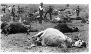
Rinderpest returned to Africa in the nineteenth century with the Italian cattle imports from Yemen and India to Somalia. The first African epidemic began in 1887, and quickly spread and killed over 90% of the domestic and wild ruminant populations in Ethiopia, Kenya, the Sudan, and Uganda leading to famines leaving majority of Ethiopia’s population dead. The disease traveled at fast pace, reaching Cape of Good Hope in 1897, finally burning itself out in 1905, all attempts to manage it having failed [2]. Many East African tribes, predominantly the Maasai, rely heavily on their cattle as a source of food, material, money, and status – a vital element of their culture. The sweeping epidemic crippled them, and eased the colonization and control of several East African countries by Europeans. Rinderpest also infects water buffalo, yaks, African buffalo, giraffes, elands, kudus, wildebeest, and Tragelaphine species of antelopes [8] impacting ecosystems as well as human societies. The wildlife acted as a reservoir for the virus, not present in Europe, which also was being increasingly urbanized. This allowed the virus to persist for longer in Africa, and failed to resurface in Europe with its historical intensity after the end of the 19th century.
European cattle imports brought rinderpest to Indonesia and the Philippines during the countries’ colonization in the late nineteenth century. The resulting epidemic spread into mainland Asia and killed 90% of the livestock in the affected areas. It burnt out and was eliminated by the veterinary programs after thirty years [2]. There is historical evidence that rinderpest depleted cattle in India and other Middle Eastern countries, where it remained endemic, several thousand years ago, but never caused the widespread ruin there as it did to Europe and Africa in recent centuries [2]. Mountain ranges and other geographic barriers likely prevented rinderpest from spreading to the rest of Asia from these regions.
The virus never established across the Atlantic Ocean, or in Australia, despite the cattle imports there. [2]
Characteristics
Rinderpest virus (RPV) has been categorized as a Morbillivirus, a genus of Paramyxoviruses. It is closely related to the measles, canine distemper virus of dogs and big cats, peste des petits ruminants virus (PPR) of shoat (goats and sheep), and the phocine distemper virus of seals. Despite its close relation to the measles, rinderpest has no effect on humans and is not dangerous to handle. The virus does have the ability to sometimes asymptomatically infect dogs and shoat, which can then carry and shed the virus [2].
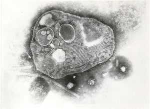
The virion ranges from 1200 to 3000 angstroms in diameter. It has a spherical capsid and inner envelope with surface proteins projecting about 90 angstroms off of the surface [6]. It has a nonsegmented, negative sense single stranded RNA genome 15,881 base pairs long. Its genome codes for eight proteins transcribed from a single promoter at the 3 prime end of the genome: six structural (large, phosphoprotein, hemagglutinin, nucleoprotein, fusion, and membrane proteins [1] and two non-structural, the functions of which are still unclear. Three distinct lineages have been defined, aptly named 1, 2, and 3. Lineages 1 and 2 are the African strains, and Lineage 3 is the Asiatic [8]. There is no available genetic material from the European epidemics to analyze for descent.
During an infection, the virus’ surface proteins bind to receptors on susceptible cells, and the virion then fuses with the host cell, emptying its genetic content and own polymerase. The polymerase then immediately begins to transcribe the genome from a single promoter, forming a leader RNA and six mRNAs for each structural protein [7]. A host nuclear protein, La protein, interacts with the leader RNA and might be involved in inducing viral RNA replication. It usually resides within the nucleus, but has been found in the cytoplasm in infected cells, implicating its involvement. The full process is currently not fully understood [7]. Host ribosomes then translate the viral mRNAs and the newly assembled virions bud off to infect more cells. The virus does not have a carrier phase.
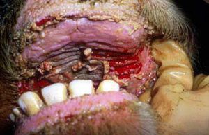
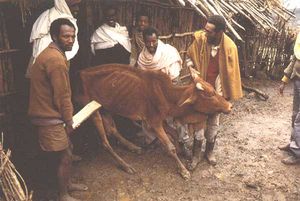
The virus moves quickly. It can be transmitted via direct contact, contaminated water, and through short distances in aerosolized bodily fluids through the air. It is not capable of vertical transmission [8]. The virion can be easily deactivated by desiccation from heat and sunlight, with a half life of two to three hours at body temperature and less than four minutes at 132 degrees Fahrenheit [6]. However the close packing and herding of domestic cattle and the normal behavior of most the affected ruminants to herd, with their own and other species, aid in the virus’ transmission. The incubation lasts less than three weeks, with symptoms arising as soon as three days after infection. The virus targets the lymphatic system, and the epithelial cells in the respiratory system and gastrointestinal tract [8]. Early signs include high but short-term fever, depression, loss of appetite, and soreness along the gums. Nasal and ocular discharge follows, then by necrotic lesions in the mouth and gastrointestinal tract, bloody diarrhea, hemorrhaging, swollen lymph nodes, and dehydration. The animal then dies as early as ten days after initial infection [8]. An animal is contagious a few days before nasal and ocular discharge appears and all bodily fluids are infectious. During an epidemic, morbidity and mortality is between 80 to 100 percent each.
Rinderpest is often collected from the gums, lymph, and spleen tissues of animals. It can be cultured in cow leukocyte and heparin (as an anticoagulant) serum and kept cold, but unfrozen [8]. Unprocessed samples may be frozen for transport. To isolate the virus from a blood sample, the sample is centrifuged to separate out the plasma and the red blood cells. The plasma is then removed, mixed with saline and re-centrifuged to remove antibodies. The resulting pellet can then be cultured on a monolayer of calf kidney cells. The virions can be identified using fluorescence-marked antibodies [8].
Vaccine Development
Before a vaccine was developed, quarantine and euthanasia were the best solutions for controlling an outbreak. The virus moved too quickly through populations for any effective treatment to be developed. In the mid-nineteenth century, farmers attempted to disinfect the remaining carcasses and control the spread using carbonic acid and burning. It was found that in the unlikely event that a cow survived, it remained resistant for the rest of its life. Early attempts at immunization started in the 1890s included injecting cattle with blood from an infected or recovered animal [10], but this ran the risk of causing an infection. In a breakthrough in 1920, J.T. Edwards infected goats, which harbor the virus without symptoms due to its close similarities with the peste-des-petis ruminants that infect shoat. After 600 passages, the virus had mutated to have reduced symptoms in cattle and could be used for immunization without killing them [2].
Plowright Tissue Culture Vaccine
Walter Plowright developed the tissue culture rinderpest vaccine (TCRV), also called the Plowright tissue culture vaccine (PTCV), in 1962. Plowright harvested samples of the Kabete “O” RPV, the most virulent strain of RPV, from the gums of infected animals and adapted it for culture in single layers of calf kidney cells [11]. After ninety or more passages through the cells, the resulting virus had become completely nonpathogenic, and could be used for inoculation [11]. The exact methods from his original paper published 1962 could not be found for the purposes of this article. Plowright’s are classic methods of formulating a live attenuated viral vaccine. RPV does not typically infect kidney tissue, but through repeat culturing in the medium, all virions in the new strain will target solely that target with reduced virulence. Exposure to the new strain prompts a rapid immune response before symptoms can begin, and the immune system’s memory cells remain afterwards. Also, although the virus has an RNA genome, which typically mutate too quickly for an effective long-term vaccine, the RPV serotype, its surface proteins, has remained the same, even through the attenuation process. This trait means that a vaccine for one RPV strain is effective against them all, and grants life-long immunity.
Plowright continued his research for many years following, investigating the stability of his vaccine, the general characteristics of the virus that he could discern with the technology of the time, and the persistence of the attenuated virus in the dead meat to prevent the export of rinderpest to naïve populations. He completed most of his research at the East African Veterinary Research Organization in Kikuyu, Kenya. He received the World Food Prize in 1999 for his work, but died several months before it was officially declared completely eradicated [11].
However this vaccine has its drawbacks. The virion’s instability requires refrigeration, making it ineffective for vaccination programs in the remote, arid, rural areas where it was needed most. Cattle also sometimes displayed moderate symptoms after inoculation. A freeze-dried version of TCRV was manufactured by mixing the vaccine with several stabilizers, and drying it at -30 degrees Celsius for sixteen hours at near vacuum. The freeze-dried vaccine can be kept at 35 degrees Celsius for up to thirty days. The vaccine, however, must be used within thirty minutes of reconstitution [7], and the cold-chain sometimes failed altogether [8].
Recombinant Vaccine
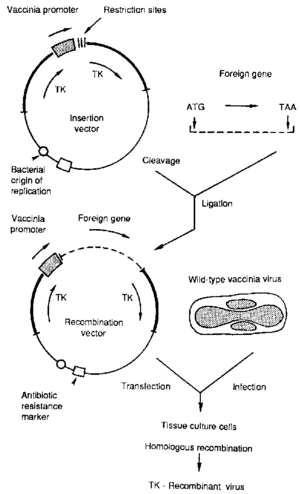
A heat-resistant recombinant vaccine was later developed in the 1980s using the virus’ surface proteins to generate an immune response. Vaccinia, the cowpox virus, is a typically asymptomatic virus with a broad host range and served as a vector for the smallpox vaccine implemented in the pathogen’s eradication. The poxvirus has the ability to incorporate heterologous genes into its genome and to express them faithfully, making it a prime vector for recombinant vaccines. Vaccinia has been designed to express RPV hemagglutinin (HA) and/or fusion (F) surface proteins to induce protective immunity. The hemogglutinin surface protein binds to host cell receptors and the fusion protein then aids in the penetration and spread of the virus to other cells [1]. Most vaccinia RPV vaccines use only the F surface protein, as it tends to elicit a stronger immune response and to be the most conserved [3], but several vaccines use both for maximum efficiency. The mRNAs coding for the surface proteins first had to be cloned and synthesized as complementary DNA, as vaccinia has a double stranded DNA genome. Plasmid vectors and transferase derived from E. coli then direct the cloning of these genes into the vaccinia genome. The genes for the RPV surface proteins are spliced using the transferase onto a vaccinia plasmid, replacing several of its genes. This transfer plasmid is then introduced into a culture infected with normal vaccinia, which uptake the plasmid via transformation (Figure 5). The resulting recombinant vaccine provides life-long immunity equivalent to that of the tissue culture vaccine with only one inoculation [14]. Furthermore, this vaccine has potential for immunizations against the peste des petits ruminants virus, which still infects sheep and goats [5]. Currently, only attenuated rinderpest and PPR vaccines are available for providing temporary protection for three to four years against PPR [14].
Because vaccinia is completely heat-resistant, transport and storage of the vaccines are no longer an issue, making it accessible in developing areas. Another advantage of the recombinant vaccine over the live attenuated one, is that inoculated animals can be differentiated from naturally infected individuals, because of vaccinia’s separate markers. This aids in disease diagnosis and control, and prevents the banning of importing vaccinated animals [9].
Efficacy
The success of both vaccines has been tested again and again, with multiple challenges of deadly doses of RPV. One described experiment injected cattle with RPV thirty-five days after inoculation into a lymph node region for additional insurance that the virus will propagate. All control animals of the study had died by day six, but no symptoms were observed after two weeks in the protected group. The vaccine did create several lesions at the injection sites, which healed two weeks later with no other symptoms [15]. In another trial, the recombinant produced similar lesions and mild fever, both of which also eventually disappeared [9]. Subsequent work on the recombinant vaccine by increasing the expression of the surface proteins resulted in products that did not give any side effects [1] [14].
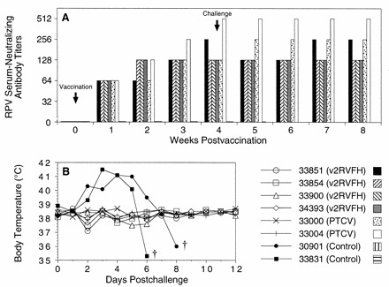
The second-generation recombinant vaccine (v2RVFH) provides the same level of protection as TCRV [14]. V2RVFH expresses large numbers of both F and H surface proteins, inducing a better and faster immune response. In a comparative study [14], two cattle were inoculated with TCRV and four with the new recombinant vaccine. Two animals were left unvaccinated as a control. None of the cattle reacted negatively to the vaccinations. The antibody levels and body temperatures were monitored throughout the process (Figure 6). By the third week, all of the inoculated cattle had built up a protective number of antibodies in their systems. Four weeks later, all of the cattle were challenged with an injection of RPV. The antibody level and body temperature remained constant in the immune cattle. The control individuals, however, spiked fevers by the third day, never produced antibodies, and both were dead by the eighth day. This vaccine was also found to provide sterilizing immunity, meaning the virions all died in the host animals, as they were unable to replicate. Virions could be isolated from nasal swabs from the controls, but not from the immune cattle, as early as three days after exposure. This means that vaccinated animals will not asymptomatically carry and transmit the virus to naïve individuals.
Eradication
Multiple influential organizations arose to combat cattle plague. The first veterinary school opened in 1762 in Lyons, France to educate its students on controlling the disease. The Office International des Epizooties, or the World Organization for Animal Health, was founded in 1924 to circumvent the difficulties of acting across international borders to battle rinderpest. The Inter-African Bureau of Epizootic Diseases began in the first efforts to completely eradicate the virus in Africa in 1950. It enjoyed initial success using the early vaccines, but it ultimately failed to maintain its inertia, mainly due to complacency in the vaccinations and observations. The pandemic resurfaced [2].
The Pan-African Rinderpest Campaign commenced in 1986 with thirty-five participant countries spanning the east and west coasts of the continent from the Sudan to Tanzania. Not all of these involved countries reported rinderpest, but were included for border control and to take no chances. Its goal was of eradication, but also general pastoral education for improved livestock production and pasture protection. They used several mediums ranging from posters, radio, and film to educate and spread awareness to avoid the relapses of the previous programs. The campaign lead mass vaccination and surveillance programs for both domestic cattle and wildlife populations and a communication network to reach even the most remote pastoralists. Donors such as the World Bank, Office International des Epizooties, and the Food and Agriculture Organization supported this program through funding, training, advice, services, and resources [12].
While effective, this was only present in Africa, while the virus was still endemic in India and the Middle East. The Food and Agriculture Organization of the United Nations later launched the Global Rinderpest Eradication Program in 1994 with the goal of a rinderpest free world by 2010. It succeeded. The last known case of rinderpest was in 2001 in Kenya, the last vaccinations were given in 2006, and the virus was declared extinct in the wild October 2010. The entire campaign had a budget of only 3 million dollars. The Food and Agriculture Organization estimates already an increase of 70 million tons of meat and one billion tons of milk has been produced in developing countries as result [13].
Conclusion
Rinderpest ravaged Europe and Africa for centuries. It succeeded due to its short incubation time, rapid onset of acute symptoms, fast rate of spread, and the close packed herding of its targets. Rinderpest shaped much of today’s current veterinary and livestock practices and organizations. But despite its virulence, rinderpest has several conserved surface protein genes, allowing for the manufacture of effective vaccines. After a few decades of development, a heat-resistant vaccine that could be easily transported, stored, and administered in developing areas where the virus hit hardest was engineered. Finally, the most historically important animal disease has been eradicated, and already the economic benefits are being enjoyed.
References
[1] Giavedoni L., et al. 1991. A vaccinia virus double recombinant expressing the F and H genes of rinderpest virus protects cattle against rinderpest and causes no pock lesions. Proc. Natl. Acad. Sci. 88:8011-8015.
[2] History of battle against rinderpest. 2010. Food and Agriculture Organization and the International Atomic Energy Agency. United Nations.
[3] Hsu, D. et al. 1988. Cloning the fusion gene of rinderpest virus: comparative sequence analysis with other morbilliviruses. Virology 166: 149-153.
[4] Letter to the Editor. 1866. The Cattle Plague. The New York Times.
[5] Peste des Petits Ruminants. 2009. OIE Terrestrial Manual. Chapter 2.7.11.
[6] Plowright, W., J.G. Cruickshank, A.P. Waterson. 1962. The morphology of rinderpest virus. Virology. Volume 17, Issue 1:118-122.
[7] Raha, T.; Pudi, R.; Das, S.; Shaila, M.S.; “Leader RNA of Rinderpest Virus Binds Specifically With Cellular La Protein: A Possible Role in Virus Replication.” Virus Research104.2 (2004): 101-109.
[8] Rinderpest. 2008. OIE Terrestrial Manual. Chapter 2.1.15
[9] Romero, C. H. et al. 1993. Single capripoxvirus recombinant vaccine for the protection of cattle against rinderpest and lumpy skin disease. Vaccine. Volume 11, Issue 7: 737-742.
[10] Simpson, P. Feature: Eradicating Rinderpest. 2011. Cambridge University Science Magazine.
[11] Taylor W.P., W. Plowright. 1964. Studies on the pathogenesis of rinderpest in experimental cattle III. Proliferation of an attenuated strain in various tissues following subcutaneous inoculation. Journal of Hygeine 63: 263-275.
[12] Villet, Jonathan. 1998. Pan African Rinderpest Campaign Communication Guide. FAO Corporate Document Repository.
[13] Walter Plowright: developed vaccine against rinderpest. 2010. New York Times Obituaries.
[14] Verardi, P. et al. 2002. Long-Term Sterilizing Immunity to Rinderpest in Cattle Vaccinated with a Recombinant Vaccinia Virus Expressing High Levels of the Fusion and Hemagglutinin Glycoproteins. Journal of Virology. Vol. 6. No. 2. 484-491.
[15] Yilma, T., Hsu, D., Jones, L., Owens, S., Grubman, M., Mebus, C., Yamanaka, M. & Dale, B. 1988. Protection of cattle against rinderpest with infectious vaccinia virus recombinants expressing the HA or F gene. Science, 242: 1058-1061
