Citrus psorosis virus (CPsV)
A Viral Biorealm page on the family Citrus psorosis virus (CPsV)

.
Baltimore Classification
Group V: (-) sense single-stranded RNA viruses
Higher order categories
Family: Ophioviridae
Genus: Ophiovirus
Description and Significance
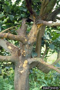
Citrus psorosis virus (CpsV) is considered to be the most infectious and problematic virus pathogen of citrus plants worldwide, however, mostly in citrus producing countries such as North, South American and the Mediterranean[1]. CpsV is the oldest reported citrus virus, first reported symptoms in 1896 and known viral etiology in 1933[2]. There are a variety of different strains and symptoms which vary from mild, type 'A,' to severe, type 'B.' External symptoms can be exhibited in leaves, fruit, bark, trunk, roots and branches. Leaf infection displays chlorotic flecks, or a decrease in chlorophyll/healthy coloring, mottling and spots; fruit may show ring-like chlorotic areas[1]. Chlorosis decreases plant's efficiency to perform photosynthesis, or ability to produce carbohydrates necessary for energy, organism support and growth[1]. The most common symptom of psorosis is deterioration of bark, such as flaking and/or scaling on trunk and branches. Early stages of this deterioration are marked by blisters or bubbles which grow to later result in bare patches[1].
Citrus plants which have been reported to exhibit this virus are grapefuit, mandarin, orange and tangerine, lemon, pomelo and lime[1]. It is economically crucial in the production and distribution of citrus fruit worldwide.
Genome Structure
Three species of ssRNA have been detected in the Citrus psorosis virus (CpsV), each of which is contained in a capsid made up of the same proteins. The three genomic negative ssRNAs are 8184, 1644 and 1454 nucleotides in length[3]. ORF 1 of RNA1, is located at 5’ end of the negative strand, and translates for a 280 kDa protein characteristic to that of RNA-dependent RNA polymerase[3]. Separated from ORF1 by 109 nucleotides is ORF2 of RNA1, which encodes a 24 kDa protein which has no homologues in the database and performs unknown functions[3]. Conversely, RNA2 has a single ORF encoding a novel protein of 54kDa which resembles a nuclear localization signal[3]. In RNA2, specifically the 3’ terminal untranslated region, there is a known polydenylation signal[3]. RNA3 has one ORF which encodes for its coat protein. The three RNAs have similar 3’ terminal regions, however, extremely differing 5’terminal regions. The particles are described as circular with a suggested “panhandle structure” which is formed by the base pairs of the terminal regions in each RNA strand; however, results vary throughout many studies of secondary structure predictions[1].
Virion Structure of CPsV virus
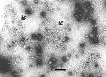
CPsV particles are characterized as spiral filaments belonging to the group of spiroviruses (SV). They are flexible and filamentous with kinks along the surface. The diameter measures about 3nm which occur in various configurations and have two known and early differentiated contour lengths of 690-760nm and the larger which is about 4 times as long. The filaments show a pattern of repeating units every 3nm along the particle length, which could possibly be coat protein molecules which form a doughnut like structure which RNA stands pass through. When in vitro, these structures which hold the RNA strands form complex linear as well as branched structures, about 9nm and half the length of the circular contour.

.
Reproductive Cycle of a CPsV virus in a Host Cell
Plant cells use a highly intricate system of exchange of RNA macromolecules, because of their complex and rigid structure; most movement is observed throughout the plasmodesmata [4]. Many components of the plant including the phloem sieve elements, vascular veins, stems and symplast, all work together to communicate and transport messages throughout the organism, including that of viral RNA[4]. In order for the CPsV to conquer intracellular spread, and shift from sites of replication throughout plant, vRNA is converted in a complex of ribonucleoprotein (vRNP). Virus-encoded pathways of transport are then carried out through infection[4].
However, little is known about ophiovirus infection in particular their localization and relationships with plant proteins [6]. Much evidence points to replication sites in the cytoplasm, in particular coat protein accumulation [6]. The coat proteins sequences show “leucine zipper motifs,” which are important in movement, capsid formation and nucleic acid binding [6].
Viral Ecology & Pathology
No confirmed transmission has been confirmed in CPsV, possible “natural” causes are reported to be the contributing factor[2]. Citrus plant areas have speculated that psorosis spreads through propagation of infected buds, soil, dodder, parasitic vines made of scales, and Olpidium-like fungus[2]. Tissue grafting has been confirmed to be one of the vectors of transmission[2].
The virus is untraceable in isolated plant sap. However, young citrus leaves exhibiting severe symptoms homogenised in 0.05 M phosphate buffer, pH 7 or Tris buffer, pH 8, both of which contain 0.5% 2-mercaptoethanol, traces of infection can be retained for at least 2 hours at 25°C or 24-48 h at 4°C [3].
CPsV ‘A’ is denoted by bark-scaling, thinning of branches, older leaves with chlorotic blotches, or pustules. Ringspots of fruits. CPsV ‘B’ causes extensive bark-scaling and branch deterioration, with chlorotic blotches in leaves appearing earlier than normal[4]. Fruits are inedible; infection is a decline in health leading to eventual death. At a molecular level, infected plant cells exhibit large quantities of abnormal cholorplasts, mitochondria and misshaping and enlargement of nuclei/nucleolous[5]. Mitochondria in particular are bent and elongated[5]. Displacement of bodies in nucleous as well as inclusion bodies which possibly contain virus particles have been reported[5].
Restoration of trees in CPsV infected areas is a rigorous process involving replanting and a bud certification program [5].
References
Example:
1) Milne, G. Robert, Garcis, M. Laura, Moreno, Pedro. Citrus psorosis virus Association of Applied Biologists. Sept, 2003. 23 Sept, 2012 <www.dpvweb.net/dpv/showadpv.php?dpvno=401> 2) Garza, Hector, da Graca, John, Prewett, Ray, Sauls, Julian, Schuster, Greta, Setamou, Mamoudou, Skaria, Mani. Citrus Greening (CG): Citrus Psorosis Virus. Citrus Texas Mutual. 23, Sept 2012. <www.valleyag.org/texascitrusgreening/diseases.php>
Page authored for BIOL 375 Virology, September 2010
