Alcaligenes faecalis NEUF2011
A Microbial Biorealm page on the genus Alcaligenes faecalis NEUF2011

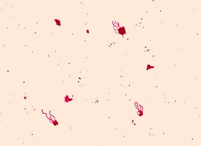
Leifson flagella stain of Alcaligenes faecalis (digitally colorized). Photograph by Dr. William A. Clark (1).
Classification
Higher order taxa
- Domain: Bacteria
- Phylum: Proteobacteria
- Class: Beta Proteobacteria
- Order: Burkholderiales
- Family: Alcaligenaceae
- Order: Burkholderiales
- Class: Beta Proteobacteria
- Phylum: Proteobacteria
Species
- Genus: Alcaligenes
- Species: faecalis
Alcaligenes faecalis
Description and significance

(a) The left eye shows edematous lids, congestion, and exudates in the anterior chamber on second postoperative day. (b) The eye on the eighth postoperative day after intravitreal injection showing reduction of exudates. (c) The eye with posterior capsule opacification at the end of second month. (d) The eye after Nd:Yag opening. Photograph by S Kaliaperumal et al (6).
Alcaligenes faecalis occur in water and soil. The microbe has peritrichous flagellar arrangement which allows for motility (2). It is a gram-negative, rod-shaped organism observed at 0.5-1.0 μm x 0.5-2.6 μm in diameter. An aerobic microbe, A. faecalis is optimal at temperatures between 20-37 °C (3).
This microbe is most commonly seen in the clinical laboratory. Most infections caused by A. faecalis are opportunistic and acquired from moist items such as nebulizers, respirators, and lavage fluids. When an infection occurs, it is usually in the form of a urinary tract infection (2). However, A. faecalis is also known to be the pathogen that causes bacterial keratitis and postoperative endophthalmitis. Numerous strains have been isolated from clinical material such as blood, urine and feces (3).
As seen in a study on metabolic energy in Alcaligenes faecalis, it was found that the microbe survives in cultures of 10 g/L of aqueous arsenic. The observed thriving of the microbe in arsenic is important in bioremediation of environments contaminated with aqueous arsenic (4). In environments with high arsenic, the community must be wary of the likely presence of A. faecalis and its tendency to cause infections.
Genome structure

Presumed gene product lengths (in amino acids [aa]) and functions are indicated. Photograph by Silver and Phung (7).
Describe the size and content of the genome. How many chromosomes? Circular or linear? Other interesting features? What is known about its sequence?
Only parts of the Alcaligenes faecalis genome have been sequenced by researchers. Silver and Phung [7] sequenced a 71-kb A. faecalis DNA region responsible for encoding arsenite oxidase as well as associated functions. More than 20 genes are located in this region, forming a "gene island" responsible for arsenic oxidation, resistance, and metabolism. These include a large molybdopterin-containing peptide subunit and a small [2Fe-2S] subunit, both responsible for arsenic oxidase.
Cell structure and metabolism
As Alcaligenes faecalis is a gram-negative bacterium, it possesses an outer membrane, a thin (compared to gram-positive bacteria) peptidoglycan layer, and a periplasm. These three layers form the gram-negative cell envelope.
Alcaligenes faecalis is unusual among gram-negative bacteria for it's ability to aerobically desaturate saturated fatty acids in order to produce monosaturated fatty acids (3). Higher-order organisms such as animals, protozoa, and various types of algae have this ability, while most bacteria and almost all gram-negative bacteria use an anaerobic pathway. Alcaligenes faecalis has also demonstrated the ability to enzymatically metabolize arsenite (AsO2-, oxidation state +3) to the less harmful arsenate (AsO4-, oxidation state +5). This bacterium could be useful for neutralization of environments contaminated by arsenite.
Ecology
Habitat; symbiosis; contributions to the environment.

The main environment in which L. acidophilus reside consists of the human stomach and intestines.(15).
Pathology
How does this organism cause disease? Human, animal, plant hosts? Virulence factors, as well as patient symptoms.
Current Research
- Crude biosurfactant from thermophilic Alcaligenes faecalis: Feasibility in petro-spill bioremediation(3)
Petroleum hydrocarbons are major environmental pollutants. Bioremediation at the site of contamination is considered an environmentally friendly means of petroleum hydrocarbon clean-up. Natural biodegradation in the environment is limited by the hydrophobic properties of hydrocarbons. Bharaliet al explored the use of A. faecallis to promote biodegradation of petroleum hydrocarbons. A. faecallisproduces biosurfactant compounds that increase the hydrophobicity of the cell surface during growth on hydrocarbons that enhances the contact with the hydrocarbons and as a result increases hydrocarbon degradation. The capacity to produce biosurfactant was studied by growing A. faecallis in salt media with a variety of hydrophobic substrates (diesel, kerosene, crude oil) as the carbon source. Under different substrate concentrations, surface tension and the rate of biosurfactant production was measured. It was found that the biosurfactant produced by A. faecallis possessed high surface activity, decreasing surface tension adequately to allow for degradation by the microorganism. In addition to the surface activity of the biosurfactant, it was found to be stable over high temperatures, a range of pH and different salt concentrations, and also possessed antimicrobial properties against a variety of bacteria and fungi. The versatile nature of the biosurfactant produced by A. faecallis makes A. faecallis an excellent candidate for use in bioremediation of hydrocarbon pollutions such as oil spills.(3)
- Improvement in ammonium removal efficiency in wastewater treatment by mixed culture of Alcaligenes faecalis No. 4 and L1(4)
Interesting Fact
Peritonitis (inflammation of the thin tissue that lines the inner wall of the abdomen and covers most of the abdominal organs) is commonly found in peritoneal dialysis (PD) patients due to contamination of the dialysis catheter. The most common pathogens causing peritonitis in PD patients are gram positive (Staphylococcus epidermidis and Staphylococcus aureus). Recently there have been unusual cases of A. faecalis causing peritonitis in PD patients. A. faecalis is not only a Gram negative bacterium but also an environmental organism. Both of these characteristics are rarely found to cause such significant infections and has since been identified as a pathogen in clinical peritonitis cases.(5)
References
2. Winn, W., Sommers, H., Koneman, E., Janda, W., Dowell, V., and Allen, S. "Color Atlas and Textbook of Diagnostic Microbiology". 'J.B. Lippincott Company'. 1988. Edition 3. p. 184, 200-201.
3. Bharali, P., Das, S., Konwar, B.K., and Thakur, A.J. (2001) Crude biosurfactant from thermophilic Alcaligenes faecalis: Feasibility in petro-spill bioremediation. Internation Biodeterioration & Biodegradation. 65:682-690.
4. Joo, H., Hirai, M., and Shoda, M. (2006) Improvement in ammonia removal efficiency in wastewater treatemnt by mixed culture of Alcaligenes faecalis No. 4 and L1. Journal of Bioscience and Bioengineering. 103(1):66-73.
5. Kahveci, A., Asicioglu, E., Tigen, E., Ari, E., Arikan, H., Odabasi, Z., and Ozener, C. (2011) Unusual causes of peritonitis in a peritoneal dialysis patient: Alcaligenes faecalis and Pantoea agglomerans. Annals of Clinical Microbiology and Antimicrobials. 10:12.
7. Simon Silver and Le T. Phung. (2005) Genes and Enzymes Involved in Bacterial Oxidation and Reduction of Inorganic Arsenic. Applied and Environmental Microbiology 71(2): 599-608.
- Created and Edited by Kevin Wieczerza, Stephanie Freed, Amanda McKenzie, Hughes Burridge -- Students of Dr. Iris Keren
