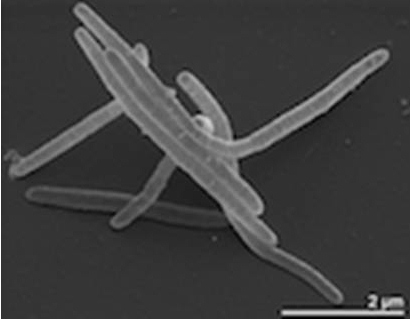
Enterococcus Avium
A Microbial Biorealm page on the genus Enterococcus Avium
Classification
Higher order taxa

Domain: Bacteria
Phylum: Firmicutes
Class: Bacilli
Order: Lactobacillales
Family: Enterococcaceae
Genus: Enterococcus
Species
E. avium
Description and significance
Enterococcus avium is a rare infection in humans, but only a few reported cases are known. In such cases, E. avium may be vancomycin-resistant Enterococcus avium, and is referred to as VREA.VREA cases have been successfully treated with linezolid.E.avium is a Gram positive, non-pigmented, non-spore forming bacterium,usually in the shape of a sphere or oval, circular, and smooth colonies.
Genome structure
Enterococcus avium contains one circular chromosome about 3445 kb in size. The sequence of 16s rRNA gene is most closely related to E. durans, with both containing six rxn operons. The whole genome has not been sequenced yet. The VREA case receives its resistance to vancomycin due to a gene called the vanA gene. There are genes that scientists have isolated from many species of enterococci (agg, gelE, ace, cylLLS, esp, cpd, fsrB) that are considered virulence factors. Analysis of the genome for E. avium indicates 35-40% G+C content of the DNA.
Cell and colony structure
Enterococcus avium is a gram positive bacterium, roughly 0.6-2.0 µm in size. It usually takes place in pairs or chains. The cells are spherically and ovoid shaped, and grow in numerous colonies consisting of thousands if not hundreds of thousands bacterium. When grown on a nutrient agar the colonies are smooth, circular and have no pigment. E. avium are non-motile bacterium . One case of VREA a women had 100,000+/mm of Enterococcus avium in her urine sample.
Metabolism
It is a strictly aerobic chemoorganotroph, meaning it uses organic compounds as its energy and carbon source. Carbon sources come from glycerol, glucose, galactose, and sucrose. Growth is observed at 10-40ºC and with 0.5–10% NaCl, with optimal growth at 28-32ºC and 4-7% NaCl.
Ecology
Marvirga tractuosa lives in a habitat that are mostly wet terrestrial habitats. They are also occasionally found in fresh water. Originally found on the beach sand from Nha Trang, Veitnam. Other strains have also been found in a variety of places: mud in the Orne Estuary, France and silty sand in Penang, Malaysia, as well as from brown mud from Muigh Inis, Ireland, underneath frozen sand in the upper littoral zone at Auke Bay, Alaska, red-brown mud from Helgoland Island, Germany, and from brown sand at Moreton Bay, Australia.
Pathology
It is not reported to be pathogenic.
References
[[2]Pagani, I.; Chertkov, O., Lapidus, A., Lucas, S., Glavina Del Rio, T., Tice, H., Copeland, A., Cheng, J., Nolan, M., Saunders, E., Pitluck, S., Held, B., Goodwin, L., Liolios, K., Ovchinnikova, G., Ivanova, N., Mavromatis, K., Pati, A., Chen, A., Palaniappan, K., Land, M., Hauser, L., Jeffries, C., Detter, J., Han, C., Tapia, R., Ngatchou, O., Rohde, M., Göker, M., Spring, S., Sikorski, J., Woyke, T., Bristow, J., Eisen, J., Markowitz, V., Hugenholtz, P., Klenk, H., & Kyrpides, N. (2011). Complete genome sequence of Marivirga tractuosa type strain (H-43T). Standards In Genomic Sciences, 4(2). doi:10.4056/sigs.1623941]
[[3]Yoon, Jaewoo, Naoya Oku, Sanghwa Park, Atsuko Katsuta, and Hiroaki Kasai. (2011) "Tunicatimonas Pelagia Gen. Nov., Sp. Nov., a Novel Representative of the Family Flammeovirgaceae Isolated from a Sea Anemone by the Differential Growth Screening Method." Antonie Van Leeuwenhoek 101.1: 133+, doi: 10.1007/s10482-011-9626-6]
[[4]Nedashkovskaya, O. I., M. Vancanneyt, S. B. Kim, and K. S. Bae. "Reclassification of Flexibacter Tractuosus (Lewin 1969) Leadbetter 1974 and 'Microscilla Sericea' Lewin 1969 in the Genus Marivirga Gen. Nov. as Marivirga Tractuosa Comb. Nov. and Marivirga Sericea Nom. Rev., Comb. Nov." International Journal Of Systematic And Evolutionary Microbiology 60.8 (2010): 1858-863. doi: 10.1099/ijs.0.016121-0]
Edited by Edited by Kayla Ferrara a student of Dr. Lisa R. Moore, University of Southern Maine, Department of Biological Sciences, [5]
