File:Cell Light Micrograph Figure 2.png
From MicrobeWiki, the student-edited microbiology resource
No higher resolution available.
Cell_Light_Micrograph_Figure_2.png (469 × 469 pixels, file size: 65 KB, MIME type: image/png)
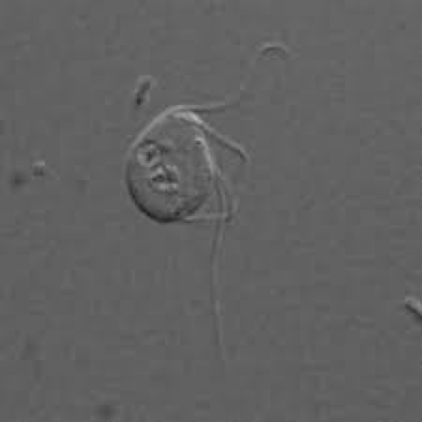
Light Micrograph of Monocercomonoides. The light micrograph images depict the grouping of flagella, which are all longer than the body, on each side of an anterior nucleus. (Google Images)
File history
Click on a date/time to view the file as it appeared at that time.
| Date/Time | Thumbnail | Dimensions | User | Comment | |
|---|---|---|---|---|---|
| current | 14:38, 12 December 2016 | | 469 × 469 (65 KB) | Slcarpen (talk | contribs) | Light Micrograph of <i>Monocercomonoides</i>. The light micrograph images depict the grouping of flagella, which are all longer than the body, on each side of an anterior nucleus. |
You cannot overwrite this file.
File usage
The following page uses this file:
