File:Geofig2.jpg: Difference between revisions
From MicrobeWiki, the student-edited microbiology resource
(geofig2.jpg.Scanning electron microscope image of bacterial cells attached to muscovite flakes within quartzite. This sample was collected from a depth of 2km in South Africa. (Photo is courtesy of M. Davidson, Princeton University, and G. Southam, Unive) |
(No difference)
|
Latest revision as of 09:11, 6 April 2011
geofig2.jpg.Scanning electron microscope image of bacterial cells attached to muscovite flakes within quartzite. This sample was collected from a depth of 2km in South Africa. (Photo is courtesy of M. Davidson, Princeton University, and G. Southam, University of Western Ontario.) [4]
File history
Click on a date/time to view the file as it appeared at that time.
| Date/Time | Thumbnail | Dimensions | User | Comment | |
|---|---|---|---|---|---|
| current | 09:11, 6 April 2011 | 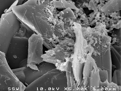 | 416 × 312 (41 KB) | Mack7 (talk | contribs) | geofig2.jpg.Scanning electron microscope image of bacterial cells attached to muscovite flakes within quartzite. This sample was collected from a depth of 2km in South Africa. (Photo is courtesy of M. Davidson, Princeton University, and G. Southam, Unive |
You cannot overwrite this file.
File usage
The following page uses this file:
