Genetics of Egg Color in Chickens: Difference between revisions
| Line 37: | Line 37: | ||
<ref>[http://www.ncbi.nlm.nih.gov/pmc/articles/PMC3847443/ Bartlett et al.: Oncolytic viruses as therapeutic cancer vaccines. Molecular Cancer 2013 12:103.]</ref> | <ref>[http://www.ncbi.nlm.nih.gov/pmc/articles/PMC3847443/ Bartlett et al.: Oncolytic viruses as therapeutic cancer vaccines. Molecular Cancer 2013 12:103.]</ref> | ||
<br><br>A citation code consists of a hyperlinked reference within "ref" begin and end codes. | <br><br>A citation code consists of a hyperlinked reference within "ref" begin and end codes. | ||
==Section 2 Microbiome== | |||
Include some current research, with a second image.<br><br> | |||
Revision as of 00:34, 28 October 2019
Introduction
Zora Mosley Chickens lay eggs in a variety of colors: white, brown, olive, green, blue, and many shades in between. The color of a chicken's egg depends on their genetic makeup. For instance, blue egg color in chickens is actually due to an ancient retrovirus that copied itself into the chicken's genome. In this article, I will be exploring the genetics behind the egg color in chickens.
Select a topic about genetics or evolution in a specific organism or ecosystem.
The topic must include one section about microbes (bacteria, viruses, fungi, or protists). This is easy because all organisms and ecosystems have microbes.
Compose a title for your page.
Type your exact title in the Search window, then press Go. The MicrobeWiki will invite you to create a new page with this title.
Open the BIOL 116 Class 2019 template page in "edit."
Copy ALL the text from the edit window.
Then go to YOUR OWN page; edit tab. PASTE into your own page, and edit.
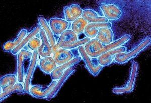
At right is a sample image insertion. It works for any image uploaded anywhere to MicrobeWiki. The insertion code consists of:
Double brackets: [[
Filename: PHIL_1181_lores.jpg
Thumbnail status: |thumb|
Pixel size: |300px|
Placement on page: |right|
Legend/credit: Electron micrograph of the Ebola Zaire virus. This was the first photo ever taken of the virus, on 10/13/1976. By Dr. F.A. Murphy, now at U.C. Davis, then at the CDC.
Closed double brackets: ]]
Other examples:
Bold
Italic
Subscript: H2O
Superscript: Fe3+
Section 1 Genetics
Include some current research, with at least one image.
Sample citations: [1]
[2]
A citation code consists of a hyperlinked reference within "ref" begin and end codes.
Section 2 Microbiome
Include some current research, with a second image.

