Human T-Lymphotropic Virus Type 1: (HTLV-1)
Introduction
This section will include an overview of the virus including history and current research
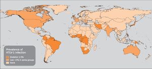
The human T-lymphotropic virus type 1 (HTLV-1) was the first oncogenic human retrovirus to be discovered. It was first studied in 1977. The virus can cause adult T-cell leukemia/lymphoma (ATL) and progressive nervous system condition known as HTLV-1-associated myelopathy or tropical spastic paraparesis (HAM/TSP)along with other neurodegenerative diseases. Although HTLV is known for being associated with lymphoma and leukemia, it more commonly causes a range of neurological disorders due to the types of cells that are infected. Human T-Lymphotropic Virus can attack a variety of cells including T cells, B cells, dendritic cells, monocytes, and endothelial cells but this virus can only edit or transform T cells and B cells. This transformation is what causes cancer in rare cases. HTLV is most commonly associated with Tropical spastic paraparesis.Tropical spastic paraparesis (TSP), is a medical condition that causes weakness, muscle spasms, and sensory disturbance resulting in weakness of the legs. However, most HTLV carriers are asymptomatic and transmit the virus to others. HTLV type 1 is less commonly associated with B cell chronic lymphocytic leukemia and T-prolymphocytic leukemia.
[1]
Structure
This section will include the structure of the virus
Human T-Lymphotropic Virus is part of the Delta-type retrovirus group. HTLVs are enveloped viruses with a diameter of approximately 80–100 nm. The HTLV virions contain two covalently bound genomic RNA strands, which are complexed with the viral enzymes reverse transcriptase, integrase and protease, and the capsid proteins. The outer part of the virions consists of a membrane-associated matrix protein and a lipid layer intersected by the envelope proteins. The genetic information is enclosed by a membrane or capsid.
[2]
Life Cycle
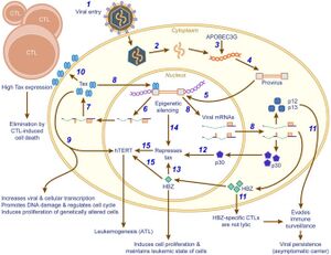
The most common type of transmission of HTLV is through blood, sexual transmission, or from mother to child via breastfeeding. After transmission Human T cell Lymphotropic Virus Type-1 is dependent on outside forces to initiate replication. A new viral cell binds to a receptor on a target cell and is integrated into the cell by a process called fusion. This begins the process of infection and replication. The virus depends on the host cell for the initial stages of transcription. In order to replicate, the viral genome must be reverse transcribed into double stranded DNA instead of RNA. The viral genome encodes for many essential genes required for the function of the virus including regulatory genes tax and rex. Tax regulates cell cycle control, increases rate of transcription, regulates cell proliferation and differentiation, and regulates DNA repair. Rex is a post transcription stabilizer and acts as an export for viral mRNAs from the nucleus to the cytoplasm.
[3]

Diagnosis
This section will include the diagnosis process of this oncogenic virus and symptoms of the infection

Conclusion
This section will include a summary
Overall text length should be at least 1,000 words (before counting references), with at least 2 images. Include at least 5 references under Reference section.
Edited by [Sydney Srnka], student of Joan Slonczewski for BIOL 116 Information in Living Systems, 2020, Kenyon College.
- ↑ (FR): IARC Working Group on the Evaluation of Carcinogenic Risks to Humans. Biological Agents. Lyon (FR):International Agency for Research on Cancer; 2012. (IARC Monographs on the Evaluation of Carcinogenic Risks to Humans, No. 100B.) "HUMAN T-CELL LYMPHOTROPIC VIRUS TYPE 1."
- ↑ Ken-ichiro Etoh, Sadahiro Tamiya, Kazunari Yamaguchi, Akihiko Okayama, Hirohito Tsubouchi, Toru Ideta, Nancy Mueller, Kiyoshi Takatsuki and Masao Matsuoka "Persistent Clonal Proliferation of Human T-lymphotropic Virus Type I-infected Cells in Vivo"
- ↑ Kannian, P., & Green, P. L. (2010) " Human T Lymphotropic Virus Type 1 (HTLV-1): Molecular Biology and Oncogenesis."
- ↑ Marco A. Lima, MD, PhD, Researcher, Instituto de e Pesquisa Clinica Evandro Chagas/FIOCRUZ, Rio de Janeiro, Brazil " HTLV Type I and Type II"
