Mycoplasma haemocanis: Difference between revisions
| Line 17: | Line 17: | ||
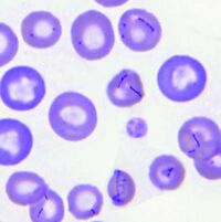
''Mycoplasma haemocanis'' is a hemotrophic, gram-negative bacterial species apart of the ''Mycoplasma'' genus. The species and genus is characterized by its lack of a cell wall, lack of flagella, and its small size of less than 1 µm. Formerly known as ''Haemobartonella canis'', this species was first discovered in Germany in 1928. It was later reclassified as a hemotrophic mycoplasma due to the similarity of its 16s ribosomal RNA (rRNA) gene and the 16s rRNA sequence of the ''Mycoplasma'' genus [4]. This species is commonly associated with causing hemolytic anemia in immunocompromised canines, specifically those who have undergone a splenectomy [1]. ''M. haemocanis'' attaches to the surface of erythrocytes (red blood cells) in hosts and can be visualized in a blood smear either as single cells or "bowed-looking" chains. This species is uncultivable ''in vitro'', and instead depends on a host cell for survival [2,4]. The bacteria has a similar resemblance to the feline equivalent ''Mycoplasma haemofelis'', which causes hemolytic anemia in cats. In a genetic comparison of the species' genomes, the two are actually concluded to be two different species infecting two distinct animals [2,3]. | ''Mycoplasma haemocanis'' is a hemotrophic, gram-negative bacterial species apart of the ''Mycoplasma'' genus. The species and genus is characterized by its lack of a cell wall, lack of flagella, and its small size of less than 1 µm. Formerly known as ''Haemobartonella canis'', this species was first discovered in Germany in 1928. It was later reclassified as a hemotrophic mycoplasma due to the similarity of its 16s ribosomal RNA (rRNA) gene and the 16s rRNA sequence of the ''Mycoplasma'' genus [4]. This species is commonly associated with causing hemolytic anemia in immunocompromised canines, specifically those who have undergone a splenectomy [1]. ''M. haemocanis'' attaches to the surface of erythrocytes (red blood cells) in hosts and can be visualized in a blood smear either as single cells or "bowed-looking" chains. This species is uncultivable ''in vitro'', and instead depends on a host cell for survival [2,4]. The bacteria has a similar resemblance to the feline equivalent ''Mycoplasma haemofelis'', which causes hemolytic anemia in cats. In a genetic comparison of the species' genomes, the two are actually concluded to be two different species infecting two distinct animals [2,3]. | ||
[[File:Mycoplasma haemocanis infection, canine blood smear - Merck Veterinary Manual.jpg| | [[File:Mycoplasma haemocanis infection, canine blood smear - Merck Veterinary Manual.jpg|200px|link=]] | ||
==Genome Structure== | ==Genome Structure== | ||
Revision as of 18:46, 14 April 2024
Classification
Bacteria; Mycoplasmatota; Mollicutes; Mycoplasmatales; Mycoplasmataceae
Species
|
NCBI: Taxonomy |
Mycoplasma haemocanis
Description and Significance
Mycoplasma haemocanis is a hemotrophic, gram-negative bacterial species apart of the Mycoplasma genus. The species and genus is characterized by its lack of a cell wall, lack of flagella, and its small size of less than 1 µm. Formerly known as Haemobartonella canis, this species was first discovered in Germany in 1928. It was later reclassified as a hemotrophic mycoplasma due to the similarity of its 16s ribosomal RNA (rRNA) gene and the 16s rRNA sequence of the Mycoplasma genus [4]. This species is commonly associated with causing hemolytic anemia in immunocompromised canines, specifically those who have undergone a splenectomy [1]. M. haemocanis attaches to the surface of erythrocytes (red blood cells) in hosts and can be visualized in a blood smear either as single cells or "bowed-looking" chains. This species is uncultivable in vitro, and instead depends on a host cell for survival [2,4]. The bacteria has a similar resemblance to the feline equivalent Mycoplasma haemofelis, which causes hemolytic anemia in cats. In a genetic comparison of the species' genomes, the two are actually concluded to be two different species infecting two distinct animals [2,3].
Genome Structure
Describe the size and content of the genome. How many chromosomes? Circular or linear? Other interesting features? What is known about its sequence?
One strain of M. haemocanis (Illinois) was isolated from the blood of an infected dog, then fully sequenced. The genome of the strain contains a singular circular chromosome comprising of 919,992 base pairs [2]. Compared to the average Mycoplasma genome size range of 580-2000 kb, the genome of M. haemocanis is consistent of species among the genus [4].
Cell Structure, Metabolism and Life Cycle
Interesting features of cell structure; how it gains energy; what important molecules it produces.
Mycoplasma haemocanis features a relatively simple cell structure.
Ecology and Pathogenesis
Habitat; symbiosis; biogeochemical significance; contributions to environment.
If relevant, how does this organism cause disease? Human, animal, plant hosts? Virulence factors, as well as patient symptoms.
References
[1] Willi, B., Novacco, M., Meli, M., Wolf-Jäckel, G., Boretti, F., Wengi, N., Lutz, H., & Hofmann-Lehmann, R. (2010). Haemotropic mycoplasmas of cats and dogs: transmission, diagnosis, prevalence and importance in Europe. Schweizer Archiv fur Tierheilkunde, 152(5), 237–244. https://doi.org/10.1024/0036-7281/a000055
[2] do Nascimento, N.C., Santos, A.P., Guimaraes, A.M. et al. (2012). Mycoplasma haemocanis – the canine hemoplasma and its feline counterpart in the genomic era. Veterinary Research, 43(1), 66. https://doi.org/10.1186/1297-9716-43-66
[3] Hemotropic Mycoplasma infections in animals - circulatory system. Merck Veterinary Manual. (n.d.). https://www.merckvetmanual.com/circulatory-system/blood-parasites/hemotropic-mycoplasma-infections-in-animals
[4] Messick, J. B. (2004). Hemotrophic mycoplasmas (hemoplasmas): A review and new insights into pathogenic potential. Veterinary Clinical Pathology, 33(1), 2–13. https://doi.org/10.1111/j.1939-165x.2004.tb00342.x
Author
Page authored by Erin Byers & Olivia Choros, students of Prof. Jay Lennon at IndianaUniversity.
