Ocular Infection by Chlamydia trachomatis: Public Health Responses to Pathology: Difference between revisions
| Line 3: | Line 3: | ||
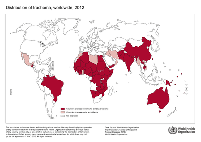
[[Image:Trachoma 2012 WHO .png|thumb|400px|right|The global distribution of ocular trachoma in 2012. Countries and geographic areas endemic for blinding trachoma shown in dark red, and areas under surveillance by the World Health Organization shown in pink. Image courtesy of WHO 2013. [http://gamapserver.who.int/mapLibrary/Files/Maps/Trachoma_2012.png ].]] | [[Image:Trachoma 2012 WHO .png|thumb|400px|right|The global distribution of ocular trachoma in 2012. Countries and geographic areas endemic for blinding trachoma shown in dark red, and areas under surveillance by the World Health Organization shown in pink. Image courtesy of WHO 2013. [http://gamapserver.who.int/mapLibrary/Files/Maps/Trachoma_2012.png ].]] | ||
<br>By Lydia Wolf<br> | <br>By Lydia Wolf<br> | ||
<br> The World Health Organization estimates ocular infection by the organism Chlamydia trachomatis to affect approximately 1.8 million people worldwide (WHO 2015). Repeated infection by the intracellular parasite, which typically begins during childhood, causes inflammation of the skin epithelium lining the inside of the eyelids called the conjunctiva. The chronic infection and inflammation of the conjunctiva causes the eyelashes of the upper lid to turn in and rub directly against the surface of the cornea. This rubbing against the cornea leads to scarring and, when left untreated by a trachiasis surgical procedure, ultimately leads to blindness…making it the leading cause of infectious blindness worldwide (Kuper, Solomon, Buchan, et al. 2003). <br> | |||
<br>At right is a sample image insertion. It works for any image uploaded anywhere to MicrobeWiki.<br><br>The insertion code consists of: | <br>At right is a sample image insertion. It works for any image uploaded anywhere to MicrobeWiki.<br><br>The insertion code consists of: | ||
<br><b>Double brackets:</b> [[ | <br><b>Double brackets:</b> [[ | ||
Revision as of 02:27, 27 April 2016
Introduction and Significance

By Lydia Wolf
The World Health Organization estimates ocular infection by the organism Chlamydia trachomatis to affect approximately 1.8 million people worldwide (WHO 2015). Repeated infection by the intracellular parasite, which typically begins during childhood, causes inflammation of the skin epithelium lining the inside of the eyelids called the conjunctiva. The chronic infection and inflammation of the conjunctiva causes the eyelashes of the upper lid to turn in and rub directly against the surface of the cornea. This rubbing against the cornea leads to scarring and, when left untreated by a trachiasis surgical procedure, ultimately leads to blindness…making it the leading cause of infectious blindness worldwide (Kuper, Solomon, Buchan, et al. 2003).
At right is a sample image insertion. It works for any image uploaded anywhere to MicrobeWiki.
The insertion code consists of:
Double brackets: [[
Filename: PHIL_1181_lores.jpg
Thumbnail status: |thumb|
Pixel size: |400px|
Placement on page: |right|
Legend/credit: World Health Organization. (2013). World Health Organization Map Production: Control of Neglected Tropical Diseases (NTD). [5].
Closed double brackets: ]]
Other examples:
Bold
Italic
Subscript: H2O
Superscript: Fe3+
Introduce the topic of your paper. What is your research question? What experiments have addressed your question? Applications for medicine and/or environment?
Sample citations: [1]
[2]
A citation code consists of a hyperlinked reference within "ref" begin and end codes.
Section 1

Include some current research, with at least one figure showing data.
Every point of information REQUIRES CITATION using the citation tool shown above.
Section 2
Include some current research, with at least one figure showing data.
Section 3

Include some current research, with at least one figure showing data.
Section 4

Conclusion
References
Authored for BIOL 238 Microbiology, taught by Joan Slonczewski, 2016, Kenyon College.
