Ophiostoma novo-ulmi: Difference between revisions
| Line 12: | Line 12: | ||
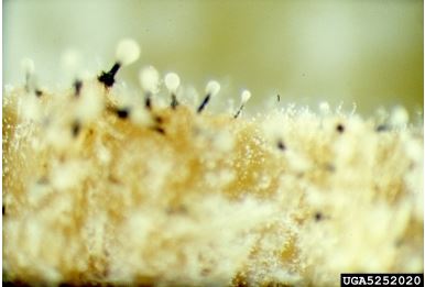
[[Image:ophiostoma.jpg|thumb|400px|right|Ophiostoma Novo-Ulmi fruiting bodies that form in galleries made by the bark beetle. Photo: Joseph OBrien, USDA Forest Service, Bugwood.org http://www.invasive.org/browse/detail.cfm?imgnum=5252020.]] | [[Image:ophiostoma.jpg|thumb|400px|right|Ophiostoma Novo-Ulmi fruiting bodies that form in galleries made by the bark beetle. Photo: Joseph OBrien, USDA Forest Service, Bugwood.org http://www.invasive.org/browse/detail.cfm?imgnum=5252020.]] | ||
=2. Introduction= | =2. '''Introduction'''= | ||
''Ophiostoma novo-ulmi'' is classified as an ascomycete fungus that spreads its spores by projecting them from elongated sacs (asci) into the air. Morphologically it consists of white/grey hyphae and a perithecial neck (250-640 µm in width), which encloses the asci. Also ''O. novo-ulmi'' can withstand temperatures of up to 33°C and the fungi’s optimal growth temperature is around 20-22°C [[#References|[2]]]. | ''Ophiostoma novo-ulmi'' is classified as an ascomycete fungus that spreads its spores by projecting them from elongated sacs (asci) into the air. Morphologically it consists of white/grey hyphae and a perithecial neck (250-640 µm in width), which encloses the asci. Also ''O. novo-ulmi'' can withstand temperatures of up to 33°C and the fungi’s optimal growth temperature is around 20-22°C [[#References|[2]]]. | ||
In the 1960s ''O. novo-ulmi'' was found to cause Dutch Elm Disease (DED), which devastated the elm tree population throughout North America [[#References|[3]]]. The pathogenicity of ''O. novo-ulmi'' can be attributed to the Pat1 gene, as well as metabolic products like diaporthic acid and phenolic acids, which are toxic to elm tree species [[#References |[4]]] [[#References |[5]]] [[#References |[6]]. Through thorough research, the mechanisms and cellular components of ''O. novo-ulmi’s'' pathogenicity seem to be very well understood, but there is more research needed on the other processes unrelated to the pathogenicity. | In the 1960s ''O. novo-ulmi'' was found to cause Dutch Elm Disease (DED), which devastated the elm tree population throughout North America [[#References|[3]]]. The pathogenicity of ''O. novo-ulmi'' can be attributed to the Pat1 gene, as well as metabolic products like diaporthic acid and phenolic acids, which are toxic to elm tree species [[#References |[4]]] [[#References |[5]]] [[#References |[6]]. Through thorough research, the mechanisms and cellular components of ''O. novo-ulmi’s'' pathogenicity seem to be very well understood, but there is more research needed on the other processes unrelated to the pathogenicity. | ||
Revision as of 02:25, 26 May 2016
1. Classification
Higher order taxa
Eukaryota; Opisthokonta; Fungi; Dikarya; Ascomycota; saccharomyceta; Pezizomycotina; leotiomyceta; sordariomyceta; Sordariomycetes; Sordariomycetidae; Ophiostomatales; Ophiostomataceae; Ophiostoma
Species
|
NCBI: [1] |
Ophiostoma novo-ulmi subsp. americana
Ophiostoma novo-ulmi subsp. novo-ulmi [1].

2. Introduction
Ophiostoma novo-ulmi is classified as an ascomycete fungus that spreads its spores by projecting them from elongated sacs (asci) into the air. Morphologically it consists of white/grey hyphae and a perithecial neck (250-640 µm in width), which encloses the asci. Also O. novo-ulmi can withstand temperatures of up to 33°C and the fungi’s optimal growth temperature is around 20-22°C [2]. In the 1960s O. novo-ulmi was found to cause Dutch Elm Disease (DED), which devastated the elm tree population throughout North America [3]. The pathogenicity of O. novo-ulmi can be attributed to the Pat1 gene, as well as metabolic products like diaporthic acid and phenolic acids, which are toxic to elm tree species [4] [5] [6. Through thorough research, the mechanisms and cellular components of O. novo-ulmi’s pathogenicity seem to be very well understood, but there is more research needed on the other processes unrelated to the pathogenicity. O. novo-ulmi can be further classified into two subspecies, americana and novo-ulmi, which have the ability to hybridize. This hybridization can create enhanced toxic mechanisms and host resistance that has the ability to hinder the establishment of new elm tree populations. This ultimately limits scientists’ ability to develop preventative mechanisms for population recovery [7]. There is little known about the specific differences between the subspecies, therefore this is an area open to further research. Dutch Elm Disease is spread by adult bark beetles, which carry the O. novo-ulmi spores. The spores eventually enter the secondary xylem of elm trees, which clogs the internal vessels of the tree and eventually causes death [8] [7]. Due to the aggressiveness of DED throughout Europe and North America, establishing proper control mechanisms has been a challenging feat [9]. Different solutions have been developed to protect elms from DED. For instance, as further described in the article, chemical treatments to eradicate the disease as well as genetic manipulation in order to target the root cause of pathogenicity were created [9] [10] [11]. Treating the trees with salicylic acid and genetically modifying them to resist the toxins of the fungus have been shown to be effective measures of combating the pathogen [10] [11]. Nevertheless, future research needs to be conducted to fully halt the spread of this disease.
3. Genome structure
Genomic Overview
The ascomycete fungus, O. novo-ulmi , is composed of a genome of 2617 nucleotides and has double stranded RNA [12]. The primary strand of the dsRNA contains an open reading frame (ORF) that has the potential to encode for 718 different types of proteins and the complementary strand contains two smaller ORFs that have the potential to encode for 178-182 proteins [12]. The large ORF on the primary strand contains 12 UGA codons that code for tryptophan, which terminates the translation of proteins on cytoplasmic ribosomes in the ascomycete mitochondria [12]. Two important nucleotide sequences of the O. novo-ulmi genome include the RNA-7 segment (1057 nucleotides) and the RNA-10 segment (317-330 nucleotides) [12]]. RNA-7 may be regarded as satellite DNA due to its distinct un-relatedness to any other sequence in the genome [12]. Also RNA-7 has the ability to exist as a single stranded form or as a double stranded form, while RNA-10 exists as a mosaic of sequences acquired from RNA-7 [12]. The loss of the RNA-7 and/or RNA-10 segments in the genome have been attributed to the reversion of diseased elm trees to uninfected phenotypes, thus suggesting that one or both of these segments contribute to the pathogenicity of ‘’O. novo-ulmi’’ [12].
RNA-dependent RNA polymerases
An important feature of the O. novo-ulmi genome resides in the fact that amino acid motifs are characteristic of RNA-dependent RNA polymerases (RdRps) [12]. RNA- dependent RNA polymerases are enzymes that have the ability to catalyze the replication of RNA from an RNA template. There are many conserved amino acid motifs in the O. novo-ulmi genome, which could explain O. novo-ulmi's strong pathogenic relationship to elm tree species [12]. From an important evolutionary standpoint it has been determined that the RdRps of mitochondrial dsRNAs for ascomycete fungi and basidiomycete fungi resemble that of ‘’O. novo-ulmi’’ [12]. Through multiple sequence alignments the fungal mitochondrial dsRNA-encoded RdRp-like proteins have been found to form a cluster. This explains how O. novo-ulmi is ancestrally related to the RdRps of positive-stranded RNA bacteriophages of the Leviviridae family (Enterobacteria, caulobacter, pseudomonas, and acinetobacter serve as natural hosts for these bacteriophages) [12]. Therefore, O. novo-ulmi could have gained pathogenicity through horizontal gene transfer with members of the Leviviridae family due to the link between the acquisition of disease and genomic elements like bacteriophages. However, the RdRps of other fungal RNA viruses and related plant and animal RNA viruses have also been found to be distinct from that of O. novo-ulmi [12]. Thus, further research needs to be conducted for a solid conclusion.
Transposable Elements
Transposable elements (TEs) have been found to account for about 14% of the O. novo-ulmi genome [13]. TEs are repetitive sequences that have the ability to move from one chromosomal location to another creating genomic rearrangements and variability in gene expression. Three main types of transposable elements (OPHIO1, OPHIO2, OPHIO3) have been found in the genome of O. novo-ulmi [13]. All three transposons have different distribution patterns in the O. novo-ulmi genome, which could allow intraspecific hybrids to serve as genetic bridges for transposable elements [13]. Of the three, OPHIO3 has the ability to elicit gene silencing due to repeat-induced point mutations [13].
Pat1 Gene
The pathogenesis of O. novo-ulmi can be attributed to a genetic cross between the AST27 gene, a Eurasian (EAN) isolate with a low level of pathogenicity and a H327 gene, which has a high level of pathogenicity [5].The cross between these two genes has led to the conclusion that one specific gene, Pat1, has led to the pathogenicity of the O. novo-ulmi species. The Pat 1 gene is located on a 3.5 Mb chromosome and is controlled by two alleles, Pat1-h and Pat1-m, which confers high to moderate levels of pathogenicity [5]. In addition to the Pat1 gene it has also been discovered that minor genes are involved in the aggressiveness of pathogenicity, however their roles are still unknown [5].
4. Cell structure
Morphology
Colonies of O. novo-ulmi are usually grey to white colored when cultured in agar [2]. The colonies generally show mycelium that are separated into fibrous ropes [2]. The base of the fungus is spherical with numerous ostiolar (pore) hyphae [2]]. Hyphae are septated, meaning that they are divided into two separate strands [2]. Mycelium conidia, which are non-motile spores (ascospores) are also produced in abundance by ‘’O. novo-ulmi’’ [2]. Mycelial conidia aggregate into mucilaginous droplets, budding in a yeast-like fashion [2]. After prolonged cultivation, conidia often fuse to create a yeast-like mass, which leads to a waxy appearance of the colony [2].
Surface Proteins
O. novo-ulmi's pathogenicity is thought to be attributed to cerato-ulmin, a hydrophobic protein that resides on the cell surface of the fungus [6. Macromolecules like cerato-ulmin that are exposed to the cell surface are important for biological activities (spread disease) of the O. novo-ulmi fungus [6. Cerato-ulmin has been defined as a class II Hydrophobin, which is characterized as being small (75-167 amino acids), possessing an amino acid terminal sequence, localized hydrophobic regions, and eight conserved cysteine residues [6. The cerato-ulmin surface protein is important due to its ability to cause wilting and involvement in disease transmission [6. The secretion of cerato-ulmin into the xylem of elm tree species leads to an obstruction in the xylem tube, which elicits tree death [6.
5. Metabolic processes
Metabolic products of O. novo-ulmi include phenolic acids, 2-4-dihydroxyl-6-acetonylbenzoic acid, 6-(1-hydroxacetonyl) and 6-pyruval analogs [4]. These metabolic products have been found and produced on elm tissue mediums, however they are rapidly metabolized by the fungus once the carbon source of the elm tree is exhausted [4]. Of the metabolites, phenolic acids, which produce high molecular weight glycoproteins can cause wilting, but not leaf necrosis [4]. Low molecular weight glycoproteins produced by phenolic acids have shown no evidence of toxicity to elm tree species. Another metabolic product of O. novo-ulmi , diaporthic acid has been found to cause severe wilting like that of phenolic acids as well as leaf necrosis [4]. Another metabolic product found in some strains of O. novo-ulmi , 4-hydroxyphenylacetic acid has been found to be toxic to elm tree species as well as other plant species [4].
6. Ecology and Pathology
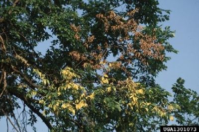
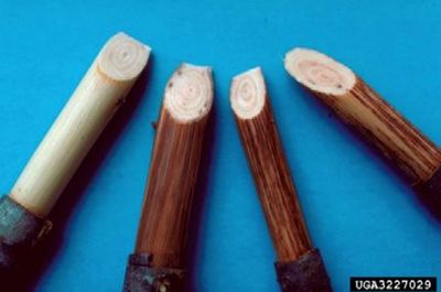
O. novo-ulmi inhabits both planted and natural forests in the Euro-American region. ‘’O. novo-ulmi’’ is the European-American strain of O. ulmi that originated in Japan [14]. This fungus survives best in cooler regions including northern Europe (originally identified in the Netherlands hence the name) and New England [13]. It can use any variety of elm trees as a host, and its relationship with the elm is parasitic while it feeds on the living tissue of the tree [14]. O. novo-ulmi will grow only in Elms and occasionally closely related plant species as most other species have a resistance to the fungus [14]. Elms often become infected when bark beetles deposit eggs in the trees’ wounds that are carrying spores of the O. novo-ulmi fungus [14].The disease spreads rapidly by the travel of beetles from diseased to healthy trees [14]. O. novo-ulmi has been found to contain an array of highly adaptive transposons that can serve as a bridge of transmission between different closely related fungi [14] (described in depth in Genome: Transposable Elements section). This contributes to its rapid spread and the devastation caused by the disease [13]. Infection with O. novo-ulmi is almost sure to cause the death of the tree once the fungus has invaded the vascular system [13]. Infection can be identified by the unseasonable wilting of leaves and brown streaking in the sapwood of wilted branches [13].
7. Disease Cycle
Young bark beetles often already carry spores of O. novo-ulmi when they emerge from pupation as the spores form in the breeding galleries of the bark beetle [15]. The beetles then move to the branches of healthy elms for maturation feeding [15]. This process most often occurs during the spring, after the beetles have made their home in already infected dead elms during the winter, and are now ready to move to new feeding grounds. As they feed on the tissue of the branches, the tree is inoculated with the O. novo-ulmi spores which then asexually reproduce through budding [2] [15].The spores can then spread through the xylem vessels of the tree and eventually cause a vascular wilt with little chances for recovery [15]. The fungus’s reproductive cycle can be continued when new beetles breed in the bark of the dying elms and continue the spread of the fungal spores to other healthy elms [15].
8. Current Research
Due to the pathogenicity of O. novo-ulmi and its ability to decimate populations of Dutch Elm trees, it is important to understand how the pathogen functions and behaves. Past and current research has been conducted in hopes of better understanding the fungus, so that therapies against O. novo-ulmi can be created to help save Dutch Elm trees. A method to estimate the rate of progression of the disease was discovered by examining sap flow measurements in Wych Elm trees inoculated with O. novo-ulmi. The first noticeable reduction in sap flow was observed six days after inoculation of Wych Elm trees and within sixteen days, the trees died. Sap flow measurement is an effective tool to study the progression of the disease in inoculated trees because it is a non-invasive method that can be used to continuously, monitor quantitatively, the development of the disease in elm trees [10]. Fungal pathogens are often tested with chemical elicitors like the endogenous plant hormone salicylic acid (SA) and simple phenols, like the essential oil component carvacrol (CA), to see if their virulence is reduced. In previous studies, these treatments were shown to significantly reduce O. novo-ulmi symptoms in elm trees. In a current published study, the chemical changes caused by SA and CA in elms were studied to better understand how these compounds reduce O. novo-ulmi symptoms in elm trees [16]. The study indicated that elm trees exhibit different metabolic responses to treatment with SA and CA. One example of this difference was that SA treated trees had an accumulation of sinapyl alcohol, while this did not occur in CA treated trees. Also, 600 mgL-1 treatments of both compounds provided similar protection for both trees, however only SA treatment improved by 12.6% from 400 to 600 mgL-1 [16]. Increase in sinapyl alcohol in SA treatment is a significant finding as the compound is a precursor for lignin, that is biosynthesized via the phenylpropanoid pathway. It was previously reported that in elm calli trees, activation of phenylpropanoid metabolism was a defense response to ‘’O. novo- ulmi’’ infection and its activation was determined to be important in the tree’s ability to suppress the pathogen. This indicates that using SA as a treatment during the early stages of infection by ‘’O. novo- ulmi’’ may improve elm tree resistance to the pathogen [16]. While treatments to suppress O. novo-ulmi have been continuously studied, extensive research and examination of various Ulmus minor trees in Spain has enabled the Spanish Environmental Administration to register seven Ulmus minor trees as tolerant to the pathogen. After 27 years of selecting and breeding for genotypes tolerant to the disease, this program has worked to increase the viability of native elm trees against the disease. The program was modeled off of a concept used in countries such as the Netherlands, where Asian elms that are resistant to O. novo-ulmi are crossed with native elms of a particular region [11]. Through this crossing, a range of hybrid clones with different genetic backgrounds and tolerance levels are created. In Spain, the law prohibited the use of crossing between foreign, tolerant Asian elms and native Spanish elms, so native Spanish elm trees were studied for resistance over 27 years. With roughly 0.5% of native elm trees showing some level of tolerance to the pathogen, the process of conserving and breeding Spanish elms that are tolerant to the pathogen is a long process, but has resulted in seven Ulmus minor trees that are tolerant to the pathogen [11]. This study is significant as it is a promising discovery in the effort to conserve the Dutch Elm Tree species.
8. References
[1] National Center for Biology Information.Web. 30 Oct. 2015.
[9] http://www.sisef.it/iforest/contents/?id=ifor1211-008 Bernier L,Mirella Aoun, Guillaume F Bouvet, André Comeau, Josée Dufour, Erika S Naruzawa, Martha Nigg, Karine V Plourde. 2014. Genomics of the Dutch elm disease pathosystem: are we there yet? iForest. 8 149-157.]
[10]) Martin, Juan A., Alejandro Solla, Maria C. Garcia-Vallejo, and Luis Gil. 2014. “Chemical changes in Ulmus minor xylem tissue after salicylic acid or carvacrol treatments are associated with enhanced resistance to Ophiostoma novo-ulmi.” Phytochemistry 83 104-109. [11] Martin Juan A., Alejandro Solla, Martin Venturas, Carmen Collada, Jorge Dominguez, Eva Miranda, Pablo Fuentes, Margarita Buron, Salustiano Iglesias, Luis Gil. 2013. “Seven Ulmus minor clones tolerant to Ophiostoma novo-ulmi registered as forest reproductive material in Spain.” Italian Society of Silviculture and Forest Ecology 8 172-180.
[15] Ophiostoma Novo-ulmi (Dutch Elm Disease)." Ophiostoma Novo-ulmi (Dutch Elm Disease). 30 Oct 2015.
Edited by [Anna Stopa, Georgia Hutchins, Anna Jortikka, Chelsy Wood], students of Jennifer Talbot for BI 311 General Microbiology, 2015, Boston University.


