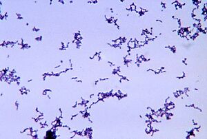
Propionibacterium acnes: A Teenager’s Worst Nightmare Defined
Section

By Megan Lydon
At right is a sample image insertion. It works for any image uploaded anywhere to MicrobeWiki.
The insertion code consists of:
Double brackets: [[
Filename: PHIL_1181_lores.jpg
Thumbnail status: |thumb|
Pixel size: |300px|
Placement on page: |right|
Legend/credit: Magnified 20,000X, this colorized scanning electron micrograph (SEM) depicts a grouping of methicillin resistant Staphylococcus aureus (MRSA) bacteria. Photo credit: CDC. Every image requires a link to the source.
Closed double brackets: ]]
Other examples:
Bold
Italic
Subscript: H2O
Superscript: Fe3+
Sample citations: [1][https://moodle.kenyon.edu/pluginfile.php/414659/mod_resource/content/0/6.%20Murphy%202021%20Spectrum.00941-21.pdf/ Gangemi, J.D.; Hightower, J.A.; Jackson, R.A.; Maher, M.H.;
Welsh, M.G.; Sigel, M.M. Enhancement of natural resistance to
influenza virus in lipopolysaccharide-responsive and -
nonresponsive mice by Propionibacterium acnes. Infect. Immun.,
1983, 39(2), 726-735.]</ref>
</ref>Bartlett et al.: Oncolytic viruses as therapeutic cancer vaccines. Molecular Cancer 2013 12:103.</ref>
A citation code consists of a hyperlinked reference within "ref" begin and end codes.
To repeat the citation for other statements, the reference needs to have a names: "Cite error: Closing </ref> missing for <ref> tag . This bacteria is typically linked to the skin condition acne vulgris, commonly known as skin acne. This species is daily commensal and highly present on healthy skin epithelium. Little is detected on the skin of adolescents, specifically those pre-pubescent. This bacterium lives on fatty acids in sebum secreted by hair sebaceous glands in hair follicles. It can also be found in the gastrointestinal biome.
Skim Microbiome
Microbiomes, in general, serve a greater purpose than living organisms just existing in their habitat. Through a combination of commensal species of microbes and their interactions with their habitat, environments are formed where the host and bacteria can adapt and regulate processes either to their advantage or negative effects of competition. The skin microbiome works identically. There is mass variability in the skin microbiome. As for microbes involved fungi, bacteria, viruses, and small arthropods contribute to this relationship [1]. In addition, the microbiome is much more complex than once thought. Past research has tended to focus only on pathogens and opportunistic pathogens rather than the entire spread of microbes in general (even “harmless” to human hosts). In addition to the variability of the microbes present in the skin microbiome, locations of the skin and their own environments are also variable from person to person. However, even in these differences, homeostasis between the microbiome and host is imperative for continued healthy interactions on the epithelium and avoids the occurrence of disease.
The skin ecosystem is continuously variable in humidity, temperature, pH, and composition of antimicrobial peptides and lipids [1]. In addition, the frequency of hair follicles can also determine the production of sebaceous materials and eccrine and apocrine glands. With this variety of environments, it establishes a separate niche for microbes to fill and thrive in. The abundance of certain bacteria is dependent on these niches.
Pathogenesis of Acnes Vulgeris by P. acnes
P acnes is present on healthy skin and disrupted skin and therefore cannot be classified as an infections disease. However, pathogenisis of P. acnes does disrupt normal epithelium and its symptoms look to be treated by many. As for it’s role in the disruption of the epethelium is the onset of acnes vulgeris. This is a condition where painful, red, and inflamed portions of the skin are infected by P. acnes. P. acnes only triggers the disease when it meets a favorable terrian, therefore the colonization of bacteria on the skin is necessary but not sufficient for pathogenesis. Research suggests that density of bacteria has no effect of frequency of acne vulgeris however there has been some evidence that certain strains of P. acnes can be more pathogenic when met with favorable conditions. However, it has been shown that there is a correlation between high sebum production and P. acnes density. Regular colonization of P. acnes is quite beneficial to the skin microbiom as it is able to hydrolyze triglycerides a release free fatty acids to maintain acidic pH in the skin surface. This then helps down regulate the density of other pathogenic bacteria such as Streptococcus pyogenes and Staphylococcus aureus.
The pilosebaceous unit is composed of three subunits: hair follicle, arrector pili muscle and sebaceous gland. The unit functions mainly as a form of protection against the external environment and aids in the dispersion of sweat. The shape of the hair follicle is also variable and can determine differences in the environments of the skin microbiome.
The transformation of the pilosebaceous unit (follicle) into the primary acne lesion is known as “Comedogensis”. During this process, P. acenes can get trapped in layers of corneocytes and excess sebum which then in turn rapidly increases colonization and presence of the bacteria in the comedonal kernel. This irregular colonization then results in the formation of a microcomedone. These microcomedomes are invisible to the naked eye but can continue to develop into a comedome. These comecomes can be a closed structure (white head) or an open condone (blackhead). Closed comedomes cannot allowed the release of cell debris, sebum and excess P. acnes and its associated products. This clog then makes the closed comdones more prone to inflammation and rupture. In this inflamed acnes vulergis, comdones rupture displacing follicular material into the dermis that than on the skin surface. The degree of damage can be classified as papules, pustules or nodules.
Substances produced by the trapped P. acnes tend to be the main reason for rupture and inflammation of closed comdones. The bacteria secrete many polypetides. Many of these polypeptides can be classified as extracellular enzymes such as proteases, hyaluronidases, neuraminidases. These polypeptides play a role in destabilizing the epithleium resulting in the possible burst and the continued formation of acnes vulgeris.
There are multiple ways in which this bacteria can harness pathogenesis of the follicular epithelium and therefore trigger an inflammatory response.
P acenes produces chemotactic factors
P. acenes produces proinflammatory cytokine inducing factors
P. acnes can activate the classical and alternative complement pathways of the innate immune system. In turn, inflammatory responses are deployed by the immune system to attack this infectious bacteria. This inflammation is mainly due to the violent neutrophils of the immune system taking in P acnes in the sebaceous follicle to release hydrolases that can then damage the follicular wall. In addition to the inflammation caused by the immune system, P acnes also releases lipases, proteases, and hyaluronidases that can add to epithelial injury.
In the toll-like receptor 2 pathway, also is important to the pathogenesis of P acnes. TLR2 is expressed on the cell surface of macrophages that are called by the immune system in acnes lesions. P acnes may trigger some sort of inflammatory cytokine response in activation once the TLR2 pathway is initiated. This interaction can lead to the upregulation of cytokine expression in sebaceous glands. However, these cytokines are also always present in these tissues even in the absence of these influences (i.e. pathogenic bacteria and inflammation).
Treatment of Acnes Vulgeris caused by P. acnes
Conclusion
References
- ↑ Cite error: Invalid
<ref>tag; no text was provided for refs namedaa
Authored for BIOL 238 Microbiology, taught by Joan Slonczewski,at Kenyon College,2024
