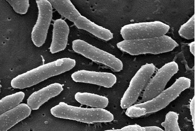
Providencia alcalifaciens
A Microbial Biorealm page on the genus Providencia alcalifaciens

Classification
Higher order taxa
Bacteria; Proteobacteria; Gammaproteobacteria; Enterobacteriales; Enterobacteriaceae; Providencia
Species
Providencia alcalifaciens
|
NCBI: [3] |
Description and significance

The definition of Abiotrophia is "life nutrition deficiency," meaning that the species needs supplemented media for growth and survival.1 Most often growing in small, satellite colonies around colonies of associated bacterial species, Abiotrophia defectiva has been shown to reside in the oral and upper respiratory flora as well as in the intestinal mucosa. It can cause bacterial, infectious endocarditis, bacteremia, and some cases of culture-negative endocarditis. 2
Genome structure
|
NCBI: Genome |
According to the NCBI database, P. alcalifaciens is made up of 4,022 protein sequences encoded in a 4.03 Mb genome. The GC content is 41.8% 2. The genome Most "P. alcalifaciens" strains are organized in plasmids. Scientists are trying to determine whether the invasiveness and genome structure of the different bacterial strains have certain correlations concerning the presence of plasmids.
Cell and colony structure
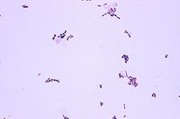
Known as a type of “nutritionally variant streptococci,” A. defectiva, when grown on blood agar, can grow as either non-hemolytic or alpha-hemolytic satellite colonies and are usually supported by many gram-positive and gram-negative bacteria. Varying from typical gram-positive streptococci to gram-variable, enlarged, pleomorphic coccobacilli, the microscopic morphology of the organisms is dependent on the type of medium. 2 When grown on 10% Danish blood agar (DBA), colonies were grayish-white in color and ranged in size from 1-3 mm in diameter. 1
Metabolism
A. defectiva is classified as a Gram-positive, non-motile, facultative aerobe. 3 A. defectiva is a fastidious organism that requires a complex medium enriched with L-cysteine or vitamin B6 as well as other unique nutritional requirements that are essential for growth. 4 Since it grows slower than other streptococci, cultivation and identification can be difficult; thus, phenotypic identification can result in a misidentification of the pathogen. 5
Ecology
PCR amplification is often used to identify A. defectiva by analyzing the 16S rDNA genes and comparing the sequence to the NCBI databank 2. A. defectiva is part of the normal flora of the oral and upper respiratory cavity as well as the intestinal tract.1
Pathology
Because A. defectiva has been frequently found in dental plaque 6, the oral cavity is often the portal of entry. 5 Although it is rare for A. defectiva to cause endocarditis, some studies estimate that it is responsible for 5-6% of all cases of inflammatory endocarditis and has a greater morbidity and mortality than endocarditis caused by other streptococci due to its poor response to many antibiotics. However, it is susceptible to and commonly treated with vancomycin.1 Complications such as congestive heart failure, embolization and an increased rate of surgical interventions often occur in conjunction with endocarditis caused by A. defectiva. The production of exopolysaccharide is one of the factors that contributes to the increased virulence of Abiotrophia species due to its long generation time which can have an impact on in vivo tolerance; the development of cell-wall deficient bacteria results in persistence, which is often promoted by treatment with β-lactam antibiotics. 5
References
(1) Christensen J and Facklam R. Granulicatella and Abiotrophia Species from Human Clinical Specimens. J. Clin. Microbiol. 2001 October; 39 [doi: 10.1128/JCM.39.10.3520-3523.2001].
(2) Hughs J, Jackson B, Kintner K, Namdari H, Namdari S, Peairs R, Savage D. Abiotrophia Species as a Cause of Endophthalmitis Following Cataract Extraction. J Clin Microbiol. 1999 May; 37(5): 1564–1566.
(3) "Abiotrophia defectiva ATCC 49176, whole genome shotgun sequencing project."
(4) http://www.ncbi.nlm.nih.gov/genome/?term=abiotrophia%20defectiva
(5) Embil J. and Vinh D (2006). Treatment of Native Valve Endocarditis: General Principles and Therapy for Specific Organisms. In K Chan & J Embil (Eds). Endocarditis: Diagnosis and Management: 121-183 [doi: 10.1007/978-1-84628-453-3_9].
(6) Beljerd M, Bouvet A, Le Coustumier A, Loubinoux J, Sire S, Wilhelm N. First case of multiple discitis and sacroiliitis due to Abiotrophia defectiva. Eur J Clin Microbiol Infect Dis. 2005; 24: 76–78. [doi: 10.1007/s10096-004-1265-7].
(7) Asma A, Mohammed A, Mushira E. Endocarditis caused by Abiotrophia defectiva. Libyan J Med. 2007; 2(1): 43–45. [doi: 10.4176/061223].
Edited by Kim Derby of Dr. Lisa R. Moore, University of Southern Maine, Department of Biological Sciences, http://www.usm.maine.edu/bio
