Section

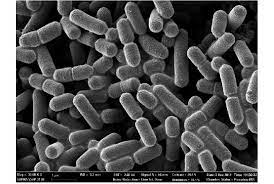
This illustration depicts a three-dimensional (3D), computer-generated image, of a group of Gram-positive, Streptococcus agalactiae (group B Streptococcus) bacteria. The photo credit for this image belongs to Alissa Eckert, who is a medical illustrator at the
CDC.
By Ryan Yarcusko
At right is a sample image insertion. It works for any image uploaded anywhere to MicrobeWiki.
The insertion code consists of:
Double brackets: [[
Filename: PHIL_1181_lores.jpg
Thumbnail status: |thumb|
Pixel size: |300px|
Placement on page: |right|
Legend/credit: Electron micrograph of the Ebola Zaire virus. This was the first photo ever taken of the virus, on 10/13/1976. By Dr. F.A. Murphy, now at U.C. Davis, then at the CDC. Every image requires a link to the source.
Closed double brackets: ]]
Other examples:
Bold
Italic
Subscript: H2O
Superscript: Fe3+
Sample citations: [1]
[2]
A citation code consists of a hyperlinked reference within "ref" begin and end codes.
To repeat the citation for other statements, the reference needs to have a names: "<ref name=aa>"
The repeated citation works like this, with a forward slash.[1]
Introduction
Include some current research, with at least one figure showing data.
Every point of information REQUIRES CITATION using the citation tool shown above.
History
Include some current research, with at least one figure showing data.

This illustration depicts a three-dimensional (3D), computer-generated image, of a group of Gram-positive, Streptococcus agalactiae (group B Streptococcus) bacteria. The photo credit for this image belongs to Alissa Eckert, who is a medical illustrator at the
CDC.
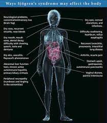
Symptoms
Include some current research, with at least one figure showing data.
This illustration depicts a three-dimensional (3D), computer-generated image, of a group of Gram-positive, Streptococcus agalactiae (group B Streptococcus) bacteria. The photo credit for this image belongs to Alissa Eckert, who is a medical illustrator at the
CDC.
Causes
This illustration depicts a three-dimensional (3D), computer-generated image, of a group of Gram-positive, Streptococcus agalactiae (group B Streptococcus) bacteria. The photo credit for this image belongs to Alissa Eckert, who is a medical illustrator at the
CDC.
Diagnosis
This illustration depicts a three-dimensional (3D), computer-generated image, of a group of Gram-positive, Streptococcus agalactiae (group B Streptococcus) bacteria. The photo credit for this image belongs to Alissa Eckert, who is a medical illustrator at the
CDC.
Treatment
Risk Factors
Complications

This illustration depicts a three-dimensional (3D), computer-generated image, of a group of Gram-positive, Streptococcus agalactiae (group B Streptococcus) bacteria. The photo credit for this image belongs to Alissa Eckert, who is a medical illustrator at the
CDC.
Microbial Consequences
Yeast Infection
edit for this image belongs to Alissa Eckert, who is a medical illustrator at the
CDC.
Dry Eyes (keratoconjunctivitis sicca)
This illustration depicts a three-dimensional (3D), computer-generated image, of a group of Gram-positive, Streptococcus agalactiae (group B Streptococcus) bacteria. The photo credit for this image belongs to Alissa Eckert, who is a medical illustrator at the
CDC.
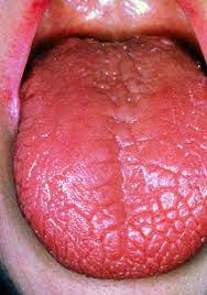
Dry Mouth (xerostomia)
This illustration depicts a three-dimensional (3D), computer-generated image, of a group of Gram-positive, Streptococcus agalactiae (group B Streptococcus) bacteria. The photo credit for this image belongs to Alissa Eckert, who is a medical illustrator at the
CDC.
Public Awareness

This illustration depicts a three-dimensional (3D), computer-generated image, of a group of Gram-positive, Streptococcus agalactiae (group B Streptococcus) bacteria. The photo credit for this image belongs to Alissa Eckert, who is a medical illustrator at the
CDC.
References
Authored for BIOL 238 Microbiology, taught by Joan Slonczewski, 2023, Kenyon College


