Solenopsis invicta virus 1: Difference between revisions
Engelbrechte (talk | contribs) |
Slonczewski (talk | contribs) |
||
| (41 intermediate revisions by one other user not shown) | |||
| Line 3: | Line 3: | ||
The imported red fire ant (<i>Solenopsis invicta</i>) has emerged as one of the most successful and destructive ant species in North America, causing an annual average of $6 billion in damages [2]. Efforts to thwart population density growth of <i>S. invicta</i> with insecticides since the 1960s have proven largely unsuccessful. In 2004, <i>S. invicta virus 1</i> was sequenced and identified as the only known viral pathogen of the imported red fire ant. Since then, SINV-2 and SINV-3 have also been identified as viruses that specifically infect <i>S. invicta</i>. Researchers are exploring how these viruses may be exploited as lethal pathogens specific to <i>S. invicta</i>. | The imported red fire ant (<i>Solenopsis invicta</i>) has emerged as one of the most successful and destructive ant species in North America, causing an annual average of $6 billion in damages [2]. Efforts to thwart population density growth of <i>S. invicta</i> with insecticides since the 1960s have proven largely unsuccessful. In 2004, <i>S. invicta virus 1</i> was sequenced and identified as the only known viral pathogen of the imported red fire ant. Since then, SINV-2 and SINV-3 have also been identified as viruses that specifically infect <i>S. invicta</i>. Researchers are exploring how these viruses may be exploited as lethal pathogens specific to <i>S. invicta</i>. | ||
[[Image:ant pic.jpg|thumb|400px|right|<i>Solenopsis invicta</i>, host of SINV-1. | [[Image:ant pic.jpg|thumb|400px|right|<i>Solenopsis invicta</i>, host of SINV-1. A fire ant's venomous sting usually causes local inflammation and a burning sensation. Although incidence is rare, anaphylaxis may result from a sting (fewer than 1% of cases).[14] ]] | ||
<br><br><br> | |||
==Virus Classification== | |||
Group IV (+) ssRNA virus, | |||
Order: Picornavirales | |||
Family:Dicistroviridae | |||
Genus: Aparavirus | |||
The dicistroviridae derive their name from the two open reading frames (cistrons) in the viral genome [5]. | |||
==Description and Significance== | ==Description and Significance== | ||
[[Image:ant ball.jpg|thumb|200px|left|<i>S. invicta</i>, Imported red fire ants adapt to inhospitable conditions. Here they have coagulated in response to increased water levels to form a raft [7]. [http://www.ars.usda.gov/IS/pr/2007/071119.htm].]] | [[Image:ant ball.jpg|thumb|200px|left|<i>S. invicta</i>, Imported red fire ants adapt to inhospitable conditions. Here they have coagulated in response to increased water levels to form a raft [7]. [http://www.ars.usda.gov/IS/pr/2007/071119.htm].]] | ||
<br>Introduced into the U.S. in the early 1900s from South America, <i>Solenopsis invicta</i>, or the red imported fire ant, is one of the most successful and invasive ant species in North America [2]. These ants exhibit uninterrupted-foraging activity, aggressive behavior, and are capable of adapting to disturbed habitats – each contributing to their ecological dominance [3]. Damage attributed to <i>S. invicta</i> includes physical damage to livestock, agricultural and electrical equipment, and human health (2, 3 | <br>Introduced into the U.S. in the early 1900s from South America, <i>Solenopsis invicta</i>, or the red imported fire ant, is one of the most successful and invasive ant species in North America [2]. These ants exhibit uninterrupted-foraging activity, aggressive behavior, and are capable of adapting to disturbed habitats – each contributing to their ecological dominance [3]. Damage attributed to <i>S. invicta</i> includes physical damage to livestock, agricultural and electrical equipment, and human health (2, 3). It has become apparent that the most effective way to control <i>S. invicta</i> population densities is through a biological mechanism. SINV-1, SINV-2, and SINV-3 are under investigation as biological control agents of the imported red fire ant [1, 2, 3, 5]. | ||
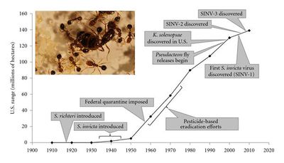
[[Image:ant graph.jpg|thumb|400px|right|<i>S. invicta</i>, Efforts to control <i>S. invicta</i> growth in the U.S. have been unsuccessful [2]. ]] | [[Image:ant graph.jpg|thumb|400px|right|<i>S. invicta</i>, Efforts to control <i>S. invicta</i> growth in the U.S. have been unsuccessful [2]. ]] | ||
<br>While a number of effective insecticides are available to control <i>S. invicta</i> population growth, they must be used regularly because of the fire ant’s ability to rapidly re-inhabit treated areas [3]. Compared to a biological mechanism of control, insecticide use is impractical from both an environmental and economic standpoint [3]. | <br>While a number of effective insecticides are available to control <i>S. invicta</i> population growth, they must be used regularly because of the fire ant’s ability to rapidly re-inhabit treated areas [3]. Compared to a biological mechanism of control, insecticide use is impractical from both an environmental and economic standpoint [3]. | ||
<br>In South America, the fire ant is exposed to more natural enemies and is rendered less invasive. These natural enemies include bacterium <i>Kneallhazia solenopsae</i>, parasitoids such as <i>Pseudacteon flies</i> and eucharitid wasps, and a parasite <i>Solenopsis daguerrei</i>. In an effort to control <i>S. invicta</i> growth, five populations of <i>Pseudacteon</i> were released in the US in the past decade [3]. Female <i>Pseudacteon</i> | <br>In South America, the fire ant is exposed to more natural enemies and is rendered less invasive. These natural enemies include bacterium <i>Kneallhazia solenopsae</i>, parasitoids such as <i>Pseudacteon flies</i> and eucharitid wasps, and a parasite <i>Solenopsis daguerrei</i>. In an effort to control <i>S. invicta</i> growth, five populations of <i>Pseudacteon</i> were released in the US in the past decade [3]. Female <i>Pseudacteon</i> launch an aerial assault down the thorax of a worker ant where they insert their eggs. 2-6 wk after oviposition, the larva migrates into the head of the fire ant and digests the tissue, ultimately decapitating the host ant [3]. Despite this lethal mechanism of parasitism, significant reductions of <i>S. invicta</i> population densities have not been observed yet [3]. First characterized in 2004, SINV-1 represented a new hope for effective, biologically-mediated extermination of <i>S. invicta</i> [1]. | ||
==Genome Structure== | ==Genome Structure== | ||
[[Image:sinv-1 genome.jpg|thumb|400px|right| Schematic diagram of the SINV-1 genome.]] | [[Image:sinv-1 genome.jpg|thumb|400px|right| Schematic diagram of the SINV-1 genome [2]. ]] | ||
The genome of SINV-1 is 8,026 nucleotides long and consists of a single (+) ssRNA (accession number AY634314) and includes a 5' UTR, two ORFs, a 3' UTR and poly A tail (as determined by Rapid Amplification of cDNA Ends) [1]. ORF1 encodes helicase, cysteine protease, and RNA-dependent RNA polymerase (RdRp). ORF2 encodes capsid proteins. SINV-1 RdRp is exhibits homology with KBV (Kashmir bee virus), Acute bee paralysis virus (ABPV), and Taura syndrome virus. The | The genome of SINV-1 is 8,026 nucleotides long and consists of a single (+) ssRNA (accession number AY634314) and includes a 5' UTR, two ORFs, a 3' UTR and poly A tail (as determined by Rapid Amplification of cDNA Ends) [1]. ORF1 encodes helicase, cysteine protease, and RNA-dependent RNA polymerase (RdRp). ORF2 is preceded by an internal ribosomal entry site and encodes capsid proteins VP1, VP2, VP3, and VP4. SINV-1 RdRp is exhibits homology with KBV (Kashmir bee virus), Acute bee paralysis virus (ABPV), and Taura syndrome virus; each are dicistroviruses [2]. The SINV-1 genome was discovered as researchers were analyzing the sequences of an expression library from an <i>S. invicta</i> colony and noticed several genes with significant homology to genes from ssRNA viruses like the Picornaviridae [1]. | ||
It has been reported that integration of (+) ssRNA viral genomes into the host genome may afford protection to the host by the corresponding virus [11, 12, 13]. Because researchers want to exploit SINV-1 as a lethal <i>S. invicta</i> pathogen, it was important to determine if the viral genome is incorporated into that of the host. In a 2004 study using 32 <i>S. invicta</i> genomic DNA samples, researchers did not detect integration of SINV-1 into the host genome, thus providing evidence of SINV-1 as a potential biopesticide [2]. | It has been reported that integration of (+) ssRNA viral genomes into the host genome may afford protection to the host by the corresponding virus [11, 12, 13]. Because researchers want to exploit SINV-1 as a lethal <i>S. invicta</i> pathogen, it was important to determine if the viral genome is incorporated into that of the host. In a 2004 study using 32 <i>S. invicta</i> genomic DNA samples, researchers did not detect integration of SINV-1 into the host genome, thus providing evidence of SINV-1 as a potential biopesticide [2]. | ||
| Line 30: | Line 35: | ||
==Virion Structure of SINV-1== | ==Virion Structure of SINV-1== | ||
<br> | <br> | ||
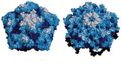
[[Image:sinv-1 capsid2.jpg|thumb|250px|right|Space-filling models of one pentamer of a Poliovirus (left) and CrPV (right) virion. Cricket paralysis virus (CrPV), like SINV-1, is a dicistrovirus with a capsid of four repeating subunits. The deep canyon surrounding the fivefold poliovirus axis is required for receptor binding [5]. ]] | |||
[[Image:SINV-1-electron.jpg|thumb|120px|left|Electron micrograph of SINV-1 particles. Scale bar represents 200 nm [2]. ]] | |||
[[Image:SINV-1-electron.jpg|thumb| | |||
The SINV-1 virion is an icosahedral, non-enveloped particle that encloses (+) ssRNA. Dicistroviruses enter their host cell via endocytosis, but it is unclear how SINV-1 specifically interacts with a receptor. The capsid is 30-31 nm in diameter and consists of four repeating viral protein (VP) subunits, VP1, VP2, VP3, and VP4 (in order of decreasing MW) [5]. | |||
==Reproductive Cycle of SINV-1 in a Host Cell== | ==Reproductive Cycle of SINV-1 in a Host Cell== | ||
| Line 51: | Line 45: | ||
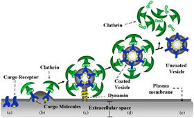
Unlike the ubiquitously replicating SINV-2 and -3, SINV-1 localizes in the gut of <i>S. invicta</i>. While the surface protein(s) that SINV-1 binds are unknown, SINV-1 likely enters its host cell via clathrin-mediated endocytosis. Once inside the cell, virions uncoat and release genomic RNA into the cytoplasm. Consistent with replication mechanisms of picornaviridae, the SINV-1 replication complex associates with virus-induced vesicles for RNA replication as a result of infection processes that remodel the Golgi apparatus [5, 9]. | Unlike the ubiquitously replicating SINV-2 and -3, SINV-1 localizes in the gut of <i>S. invicta</i>. While the surface protein(s) that SINV-1 binds are unknown, SINV-1 likely enters its host cell via clathrin-mediated endocytosis. Once inside the cell, virions uncoat and release genomic RNA into the cytoplasm. Consistent with replication mechanisms of picornaviridae, the SINV-1 replication complex associates with virus-induced vesicles for RNA replication as a result of infection processes that remodel the Golgi apparatus [5, 9]. | ||
[[Image:endocytosis1.jpg|thumb|280px|left| Dicistrovirus virions enter the host cell via clathrin-mediated endocytosis [8]. ]] | |||
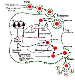
SINV-1 (+) RNA may be translated upon entry into the cytoplasm by host translational machinery (ribosomes) as a single polypeptide which is cleaved by viral proteases to produce about 7 viral proteins, including a 5’ viral protein genome (VPg, a peptide covalently linked at the 5’ terminus) which serves as a primer for viral RNA replication [2]. (+) RNA is also transcribed by SINV-1 RNA-dependent RNA polymerase to (-) RNA, which is then transcribed to (+) RNA for subsequent translation or packing into a capsid. Fully assembled SINV-1 virions spread after lysing the host cell, however it is unclear how frequently this happens as SINV-1 infected ants are typically asymptomatic [1, 2]. | SINV-1 (+) RNA may be translated upon entry into the cytoplasm by host translational machinery (ribosomes) as a single polypeptide which is cleaved by viral proteases to produce about 7 viral proteins, including a 5’ viral protein genome (VPg, a peptide covalently linked at the 5’ terminus) which serves as a primer for viral RNA replication [2]. (+) RNA is also transcribed by SINV-1 RNA-dependent RNA polymerase to (-) RNA, which is then transcribed to (+) RNA for subsequent translation or packing into a capsid. Fully assembled SINV-1 virions spread after lysing the host cell, however it is unclear how frequently this happens as SINV-1 infected ants are typically asymptomatic [1, 2]. | ||
[[Image:replication.jpg|thumb|250px|right| Canonical replication mechanism of group IV (+) ssRNA viruses [8]. ]] | |||
[[Image: | |||
==Viral Ecology & Pathology== | ==Viral Ecology & Pathology== | ||
| Line 62: | Line 54: | ||
In the laboratory, SINV-1 infection was associated with larval mortality [2]. These observations highlight the complexity of microscopic mutualisms in nature. | In the laboratory, SINV-1 infection was associated with larval mortality [2]. These observations highlight the complexity of microscopic mutualisms in nature. | ||
The goal of SINV research is to exploit these viruses so they can effectively wipe out populations of the imported red fire ant. | The goal of SINV research is to exploit these viruses so they can effectively wipe out populations of the imported red fire ant. Recent research suggests that SINV-3 may be the most lethal of the three SINV viruses. For this reason, the laboratory of Steven M. Valles is developing a SINV-3 construct and focusing their efforts on studying the production, host specificity, efficacy, dose responses, mechanism of action, and overall viability of SINV-3 as a biopesticide [2]. His laboratory hasn't discounted the potential value of SINV-1 and SINV-2 as biopesticides. His lab has proposed using these viruses to deliver a toxin or an RNAi sequence to <i>S. invicta</i> to rapidly wipe out populations of the pest [2]. | ||
==References== | ==References== | ||
| Line 85: | Line 77: | ||
[10] M. I. Hertz and S. R. Thompson, “In vivo functional analysis of the Dicistroviridae intergenic region internal ribosome entry sites,” Nucleic Acids Research, vol. 39, no. 16, pp. 7276-7288, 2011. | [10] M. I. Hertz and S. R. Thompson, “In vivo functional analysis of the Dicistroviridae intergenic region internal ribosome entry sites,” Nucleic Acids Research, vol. 39, no. 16, pp. 7276-7288, 2011. | ||
[11] E. Maori, E. Tanne, and I. Sela, “Reciprocal sequence exchange between non-retro viruses and hosts leading to the appearance of new host phenotypes,” Virology, vol. 362, no. 2, pp. 342–349, 2007. | |||
[12] E. Tanne and I. Sela, “Occurrence of a DNA sequence of a non-retro RNA virus in a host plant genome and its expression: evidence for recombination between viral and host RNAs,” Virology, vol. 332, no. 2, pp. 614–622, 2005. | |||
[13] S. Crochu, S. Cook, H. Attoui et al., “Sequences of flavivirus-related RNA viruses persist in DNA form integrated in the genome of Aedes spp. mosquitoes,” Journal of General Virology, vol. 85, no. 7, pp. 1971–1980, 2004. | |||
[14] Drees, Bastiaan M.. "MEDICAL PROBLEMS AND TREATMENT CONSIDERATIONS FOR THE RED IMPORTED FIRE ANT." . Texam A&M University Department of Entomology , 2002. | |||
<br><br> | <br><br> | ||
Page authored for [http://biology.kenyon.edu/courses/biol375/ | Page authored for [http://biology.kenyon.edu/courses/biol375/biol375syl12.html BIOL 375 Virology], September 2012 | ||
<!--Do not edit or remove this line-->[[Category:Pages edited by students of Joan Slonczewski at Kenyon College]] | <!--Do not edit or remove this line-->[[Category:Pages edited by students of Joan Slonczewski at Kenyon College]] | ||
Latest revision as of 13:26, 15 October 2012
A Viral Biorealm page on the family Solenopsis invicta virus 1
The imported red fire ant (Solenopsis invicta) has emerged as one of the most successful and destructive ant species in North America, causing an annual average of $6 billion in damages [2]. Efforts to thwart population density growth of S. invicta with insecticides since the 1960s have proven largely unsuccessful. In 2004, S. invicta virus 1 was sequenced and identified as the only known viral pathogen of the imported red fire ant. Since then, SINV-2 and SINV-3 have also been identified as viruses that specifically infect S. invicta. Researchers are exploring how these viruses may be exploited as lethal pathogens specific to S. invicta.
Virus Classification
Group IV (+) ssRNA virus,
Order: Picornavirales
Family:Dicistroviridae
Genus: Aparavirus
The dicistroviridae derive their name from the two open reading frames (cistrons) in the viral genome [5].
Description and Significance
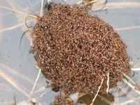
Introduced into the U.S. in the early 1900s from South America, Solenopsis invicta, or the red imported fire ant, is one of the most successful and invasive ant species in North America [2]. These ants exhibit uninterrupted-foraging activity, aggressive behavior, and are capable of adapting to disturbed habitats – each contributing to their ecological dominance [3]. Damage attributed to S. invicta includes physical damage to livestock, agricultural and electrical equipment, and human health (2, 3). It has become apparent that the most effective way to control S. invicta population densities is through a biological mechanism. SINV-1, SINV-2, and SINV-3 are under investigation as biological control agents of the imported red fire ant [1, 2, 3, 5].
While a number of effective insecticides are available to control S. invicta population growth, they must be used regularly because of the fire ant’s ability to rapidly re-inhabit treated areas [3]. Compared to a biological mechanism of control, insecticide use is impractical from both an environmental and economic standpoint [3].
In South America, the fire ant is exposed to more natural enemies and is rendered less invasive. These natural enemies include bacterium Kneallhazia solenopsae, parasitoids such as Pseudacteon flies and eucharitid wasps, and a parasite Solenopsis daguerrei. In an effort to control S. invicta growth, five populations of Pseudacteon were released in the US in the past decade [3]. Female Pseudacteon launch an aerial assault down the thorax of a worker ant where they insert their eggs. 2-6 wk after oviposition, the larva migrates into the head of the fire ant and digests the tissue, ultimately decapitating the host ant [3]. Despite this lethal mechanism of parasitism, significant reductions of S. invicta population densities have not been observed yet [3]. First characterized in 2004, SINV-1 represented a new hope for effective, biologically-mediated extermination of S. invicta [1].
Genome Structure
The genome of SINV-1 is 8,026 nucleotides long and consists of a single (+) ssRNA (accession number AY634314) and includes a 5' UTR, two ORFs, a 3' UTR and poly A tail (as determined by Rapid Amplification of cDNA Ends) [1]. ORF1 encodes helicase, cysteine protease, and RNA-dependent RNA polymerase (RdRp). ORF2 is preceded by an internal ribosomal entry site and encodes capsid proteins VP1, VP2, VP3, and VP4. SINV-1 RdRp is exhibits homology with KBV (Kashmir bee virus), Acute bee paralysis virus (ABPV), and Taura syndrome virus; each are dicistroviruses [2]. The SINV-1 genome was discovered as researchers were analyzing the sequences of an expression library from an S. invicta colony and noticed several genes with significant homology to genes from ssRNA viruses like the Picornaviridae [1].
It has been reported that integration of (+) ssRNA viral genomes into the host genome may afford protection to the host by the corresponding virus [11, 12, 13]. Because researchers want to exploit SINV-1 as a lethal S. invicta pathogen, it was important to determine if the viral genome is incorporated into that of the host. In a 2004 study using 32 S. invicta genomic DNA samples, researchers did not detect integration of SINV-1 into the host genome, thus providing evidence of SINV-1 as a potential biopesticide [2].
Virion Structure of SINV-1
The SINV-1 virion is an icosahedral, non-enveloped particle that encloses (+) ssRNA. Dicistroviruses enter their host cell via endocytosis, but it is unclear how SINV-1 specifically interacts with a receptor. The capsid is 30-31 nm in diameter and consists of four repeating viral protein (VP) subunits, VP1, VP2, VP3, and VP4 (in order of decreasing MW) [5].
Reproductive Cycle of SINV-1 in a Host Cell
Unlike the ubiquitously replicating SINV-2 and -3, SINV-1 localizes in the gut of S. invicta. While the surface protein(s) that SINV-1 binds are unknown, SINV-1 likely enters its host cell via clathrin-mediated endocytosis. Once inside the cell, virions uncoat and release genomic RNA into the cytoplasm. Consistent with replication mechanisms of picornaviridae, the SINV-1 replication complex associates with virus-induced vesicles for RNA replication as a result of infection processes that remodel the Golgi apparatus [5, 9].
SINV-1 (+) RNA may be translated upon entry into the cytoplasm by host translational machinery (ribosomes) as a single polypeptide which is cleaved by viral proteases to produce about 7 viral proteins, including a 5’ viral protein genome (VPg, a peptide covalently linked at the 5’ terminus) which serves as a primer for viral RNA replication [2]. (+) RNA is also transcribed by SINV-1 RNA-dependent RNA polymerase to (-) RNA, which is then transcribed to (+) RNA for subsequent translation or packing into a capsid. Fully assembled SINV-1 virions spread after lysing the host cell, however it is unclear how frequently this happens as SINV-1 infected ants are typically asymptomatic [1, 2].
Viral Ecology & Pathology
Late-instar S. invicta digest all solid food for the colony and redistribute it as liquid to nestmates [2]. In this way, larvae serve as both a replication site and passage fir SINV-1 colonial propagation [2]. Experiments using Real-time PCR demonstrate that SINV-1 exists primarily in the midgut of S. invicta, evidence of the horizontal fecal-oral route of viral infection [2]. SINV-1 detection in "all developmental stages of S. invicta including eggs and queen" indicates vertical transmission of the virus [2]. SINV-1 infected S. invicta populations are asymptomatic, but their ability to compete with uninfected ants of their own and other species is reduced [2]. Some environmental stresses induce symptoms or even death in SINV-1 infected ants [2]. Researchers observed that SINV-1 internal ribosome entry site within the intergenic region (IGR IRES) exhibits increased activity at higher temperatures [10]. This data correlates with an increased incidence of S. invicta SINV-1 infection at higher temperatures in field studies [2]. In the laboratory, SINV-1 infection was associated with larval mortality [2]. These observations highlight the complexity of microscopic mutualisms in nature.
The goal of SINV research is to exploit these viruses so they can effectively wipe out populations of the imported red fire ant. Recent research suggests that SINV-3 may be the most lethal of the three SINV viruses. For this reason, the laboratory of Steven M. Valles is developing a SINV-3 construct and focusing their efforts on studying the production, host specificity, efficacy, dose responses, mechanism of action, and overall viability of SINV-3 as a biopesticide [2]. His laboratory hasn't discounted the potential value of SINV-1 and SINV-2 as biopesticides. His lab has proposed using these viruses to deliver a toxin or an RNAi sequence to S. invicta to rapidly wipe out populations of the pest [2].
References
[1] Valles, Steven M., Charles A. Strong, et al. "A licorna-like virus from the red imported fire ant, Solenopsis invicta: initial discovery, genome sequence, and characterization." Virology. (2004): 151-157.
[2] Valles, Steven M. "Review Article: Positive-strand RNA viruses infecting the red imported fire ant, Solenopsis invicta ." Psyche. (2012).
[3] Briano, Juan, Luis Calcaterra, and Laura Varone. "Review Article: Fire ants (Solenopsis spp.) and their natural enemies in southern South America." Psyche. (2011).
[4] Valles, Steven M., and Yoshifumi Hashimoto. "Isolation and characterization of Solenopsis invicta virus 3, a new positive-strand RNA virus infecting the red imported fire ant, Solenopsis invicta." Virology. 388. (2009): 354-361.
[5] Bonning, Bryony C., and W. Allen Miller. "Dicistroviruses."Annu. Rev. Entomol.. (2010): 129-150.
[6] Porter, Sanford D. "Biology and behavior of pseudacteon decapitating flies (Diptera:Phoridae) that parasitize Solenopsis fire ants (Hymenoptera: Formicidai)."Florida Entomologist. 81. (1998): 292-309.
[7] United States. National Park Service. Congaree National Park. Print. <http://www.nps.gov/cong/siteindex.htm>.
[8] Sebastian, R., E. Diaz, et al. "Studying endocytosis in space and time by means of temporal Boolean models." Pattern Recognition. 39.11 (2006): 2175–2185.
[9] Belov, George A., Nihal Altan-Bonnet, et al. "Hijacking components of the cellular secretory pathway for replication of poliovirus RNA." Journal of Virology. 81.2 (2006): 558-565.
[10] M. I. Hertz and S. R. Thompson, “In vivo functional analysis of the Dicistroviridae intergenic region internal ribosome entry sites,” Nucleic Acids Research, vol. 39, no. 16, pp. 7276-7288, 2011.
[11] E. Maori, E. Tanne, and I. Sela, “Reciprocal sequence exchange between non-retro viruses and hosts leading to the appearance of new host phenotypes,” Virology, vol. 362, no. 2, pp. 342–349, 2007.
[12] E. Tanne and I. Sela, “Occurrence of a DNA sequence of a non-retro RNA virus in a host plant genome and its expression: evidence for recombination between viral and host RNAs,” Virology, vol. 332, no. 2, pp. 614–622, 2005.
[13] S. Crochu, S. Cook, H. Attoui et al., “Sequences of flavivirus-related RNA viruses persist in DNA form integrated in the genome of Aedes spp. mosquitoes,” Journal of General Virology, vol. 85, no. 7, pp. 1971–1980, 2004.
[14] Drees, Bastiaan M.. "MEDICAL PROBLEMS AND TREATMENT CONSIDERATIONS FOR THE RED IMPORTED FIRE ANT." . Texam A&M University Department of Entomology , 2002.
Page authored for BIOL 375 Virology, September 2012
