File list
From MicrobeWiki, the student-edited microbiology resource
This special page shows all uploaded files.
| Date | Name | Thumbnail | Size | User | Description | Versions |
|---|---|---|---|---|---|---|
| 00:02, 12 December 2017 | Phylogeny.jpg (file) |  |
283 KB | Siabbas | 1 | |
| 23:59, 11 December 2017 | Mycoplasmamorph.jpg (file) |  |
68 KB | Siabbas | 1 | |
| 23:55, 11 December 2017 | Mycoplasmafermentansmorphology.jpg (file) |  |
68 KB | Siabbas | 2 | |
| 22:21, 11 December 2017 | Wiki Pic.jpg (file) |  |
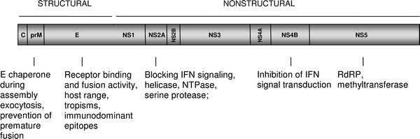
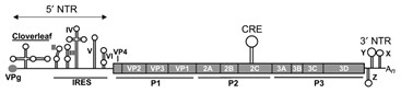
30 KB | Dtblais | Adapted and revised from Zybert (2011). Genome map of the Dengue Virus. | 1 |
| 21:47, 11 December 2017 | Rhizopus oryzae.gif (file) |  |
7 KB | Tingwat | 1 | |
| 16:38, 11 December 2017 | Mode of infection.jpg (file) |  |
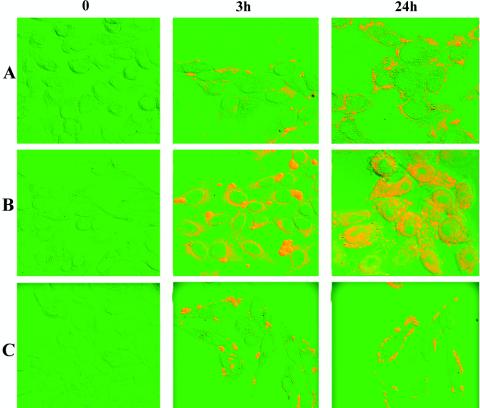
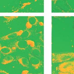
22 KB | Siabbas | Fig. 1.Confocal micrographs demonstrating binding and internalization of M. fermentans and M. pneumoniae by HeLa cells. Row A, HeLa cells infected with native M. fermentans; row B, HeLa cells infected with Pg- and uPA-treated M. fermentans; row C, HeLa... | 1 |
| 16:35, 11 December 2017 | Example.jpg (file) |  |
22 KB | Siabbas | Fig. 1 Confocal micrographs demonstrating binding and internalization of M. fermentans and M. pneumoniae by HeLa cells. Row A, HeLa cells infected with native M. fermentans; row B, HeLa cells infected with Pg- and uPA-treated M. fermentans; row C, HeLa... | 2 |
| 16:31, 11 December 2017 | Mode of infection .jpeg (file) |  |
10 KB | Siabbas | Fig.1 Confocal micrographs demonstrating binding and internalization of M. fermentans and M. pneumoniae by HeLa cells. Row A, HeLa cells infected with native M. fermentans; row B, HeLa cells infected with Pg- and uPA-treated M. fermentans; row C, HeLa... | 1 |
| 19:20, 9 December 2017 | Coxsackievirus Type B Genome.png (file) | 11 KB | Bjardin | Figure: Secondary structures of the Coxsackie Virus B RNA and the encoded proteins. Taken from van Ooij, M.J., Vogt, D.A., Paul, A., Castro, C., Kuijpers, J., van Kuppeveld, F.J., Cameron, C.E., Wimmer, E., Andino, R. and Melchers, W.J. 2006, with perm... | 1 | |
| 19:09, 9 December 2017 | Coxsackievirus Type B Intracellular Signaling.png (file) |  |
164 KB | Bjardin | Figure: CVB3-induced intracellular signalling pathways and networks in infected cells. ( a ) Attachment of CVB3 to its receptors, CAR and DAF, ( b) induces a rapid and transient activation of src-family kinases and the ERK pathway. ( c) CVB3 infectio... | 1 |
| 19:08, 9 December 2017 | Coxsackievirus Type B Proteases.png (file) |  |
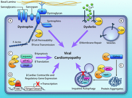
100 KB | Bjardin | Figure: Direct myocardial damage induced by virus-encoded proteinases. A, Dystrophin is an important component of the dystrophin–glycoprotein complex that links the cytoskeleton to the extracellular matrix. coxsackievirus B3–encoded proteinase 2A c... | 1 |
| 19:06, 9 December 2017 | Coxsackievirus Type B Structure.png (file) |  |
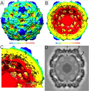
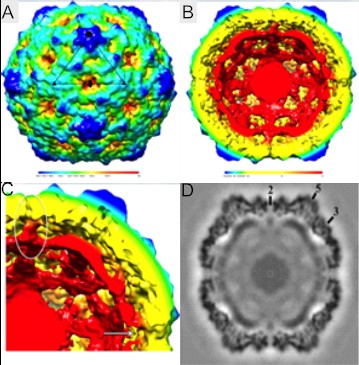
267 KB | Bjardin | Figure: CAR-catalyzed CVB3 A-particle. (A) CVB3 A-particle is shown surface rendered and colored according to radial height (key beneath map). An asymmetric unit is outlined in black. (B) Cutaway view of the A-particle showing capsid separation from ge... | 1 |
| 19:01, 9 December 2017 | Coxsackievirus Type B Structure.jpg (file) |  |
53 KB | Bjardin | Figure: CAR-catalyzed CVB3 A-particle. (A) CVB3 A-particle is shown surface rendered and colored according to radial height (key beneath map). An asymmetric unit is outlined in black. (B) Cutaway view of the A-particle showing capsid separation from ge... | 1 |
| 19:00, 9 December 2017 | Coxsackievirus Type B Genome.jpg (file) | 8 KB | Bjardin | Figure: Secondary structures of the Coxsackie Virus B RNA and the encoded proteins. Taken from van Ooij, M.J., Vogt, D.A., Paul, A., Castro, C., Kuijpers, J., van Kuppeveld, F.J., Cameron, C.E., Wimmer, E., Andino, R. and Melchers, W.J. 2006, with perm... | 1 | |
| 18:21, 9 December 2017 | Microbewiki Genome 12-9-17.png (file) | 11 KB | Bjardin | Figure: Secondary structures of the Coxsackie Virus B RNA and the encoded proteins. Taken from van Ooij, M.J., Vogt, D.A., Paul, A., Castro, C., Kuijpers, J., van Kuppeveld, F.J., Cameron, C.E., Wimmer, E., Andino, R. and Melchers, W.J. 2006, with per... | 1 | |
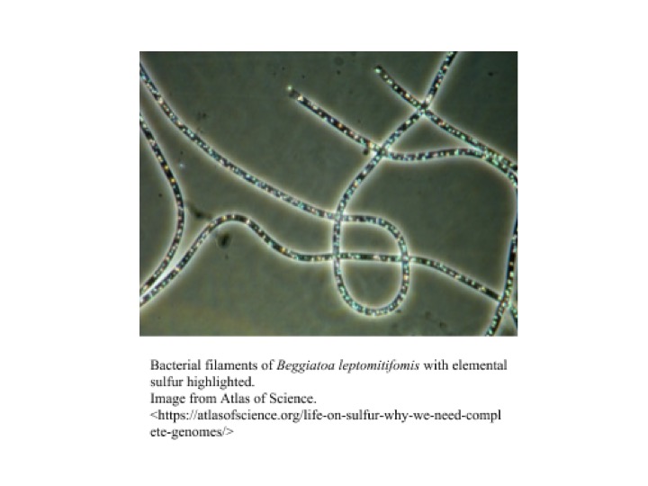
| 14:47, 8 December 2017 | Beggiatoa.jpg (file) |  |
52 KB | Ztarasie | 1 | |
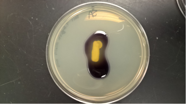
| 04:32, 8 December 2017 | Casein Hydrolisis.png (file) |  |
369 KB | Tara.eulenfeld | 1 | |
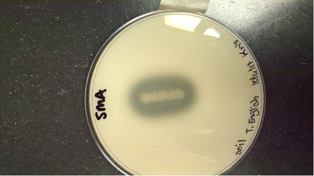
| 04:28, 8 December 2017 | Starch Hydrolysis.png (file) |  |
263 KB | Tara.eulenfeld | 1 | |
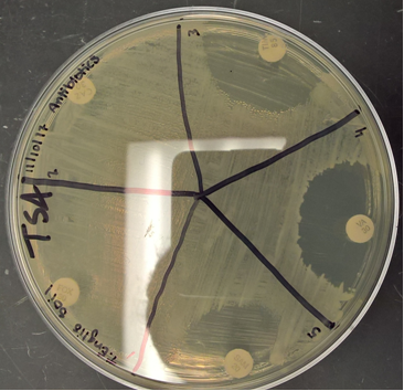
| 04:21, 8 December 2017 | Disinfectant susceptibility.png (file) |  |
236 KB | Tara.eulenfeld | 1 | |
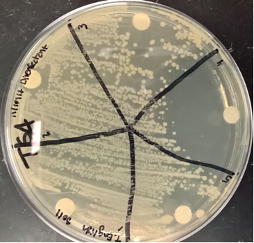
| 04:10, 8 December 2017 | Antimicrobial susceptibility.png (file) |  |
235 KB | Tara.eulenfeld | 1 | |
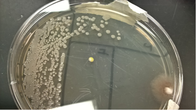
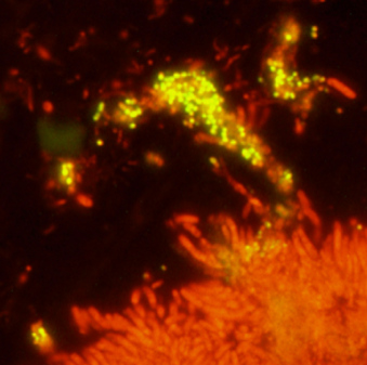
| 04:05, 8 December 2017 | Soil microorganism.png (file) |  |
331 KB | Tara.eulenfeld | 1 | |
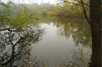
| 21:49, 7 December 2017 | Copperfield park.png (file) |  |
183 KB | Jljordan77 | 1 | |
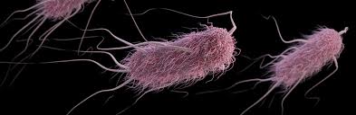
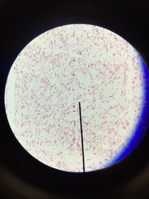
| 01:04, 7 December 2017 | GramNeg.jpg (file) |  |
33 KB | Ladell | 2 | |
| 00:47, 7 December 2017 | Eosinmethyleneblueweakpos.jpg (file) |  |
52 KB | Ladell | 1 | |
| 00:46, 7 December 2017 | Hektoenentericneg.jpg (file) |  |
58 KB | Ladell | 1 | |
| 00:44, 7 December 2017 | MacConkeyneg.jpg (file) |  |
57 KB | Ladell | 1 | |
| 00:38, 7 December 2017 | Phenylalanine deaminase neg.jpg (file) |  |
83 KB | Ladell | 1 | |
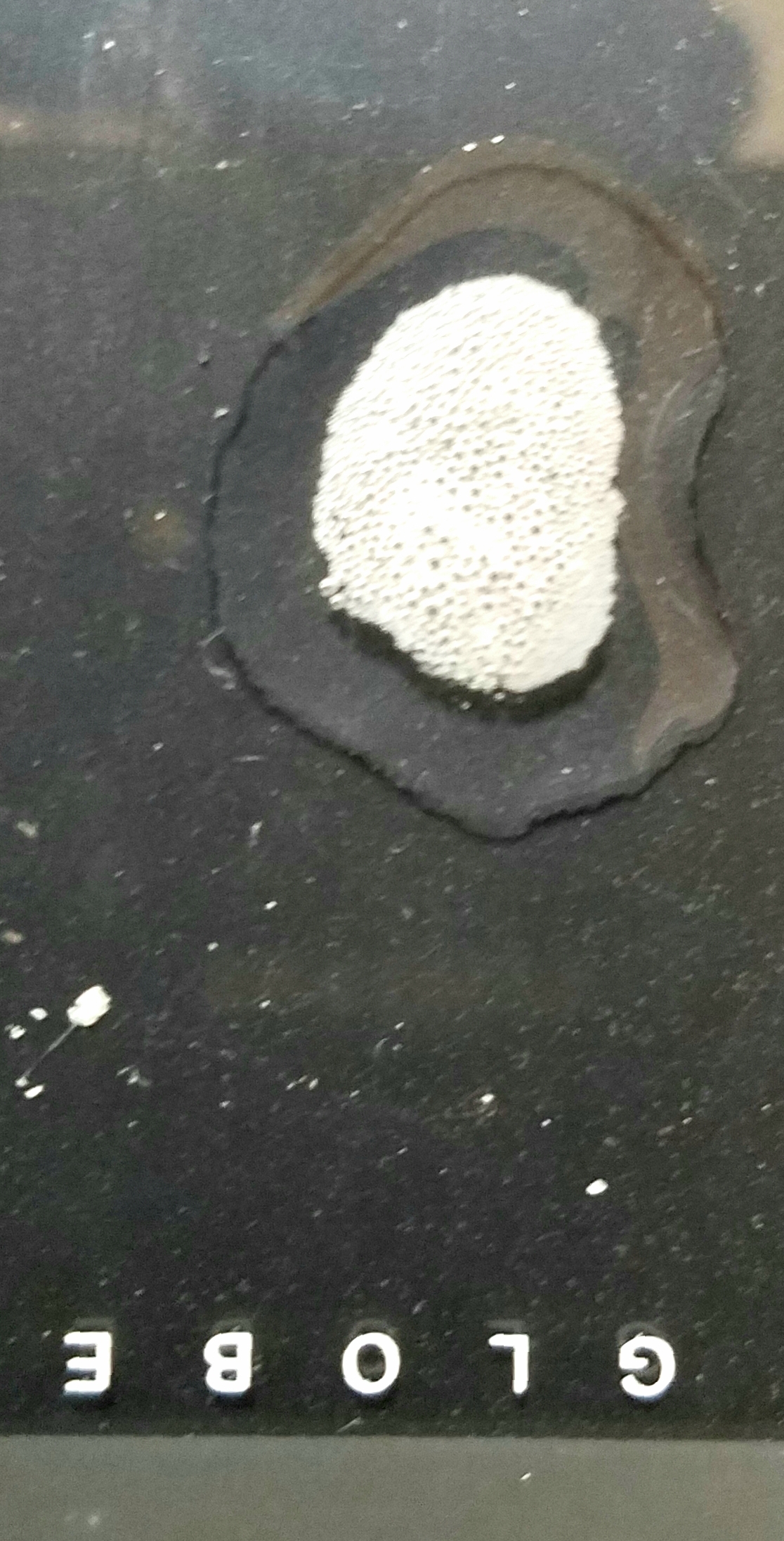
| 00:35, 7 December 2017 | Catalasepos.jpg (file) |  |
83 KB | Ladell | 1 | |
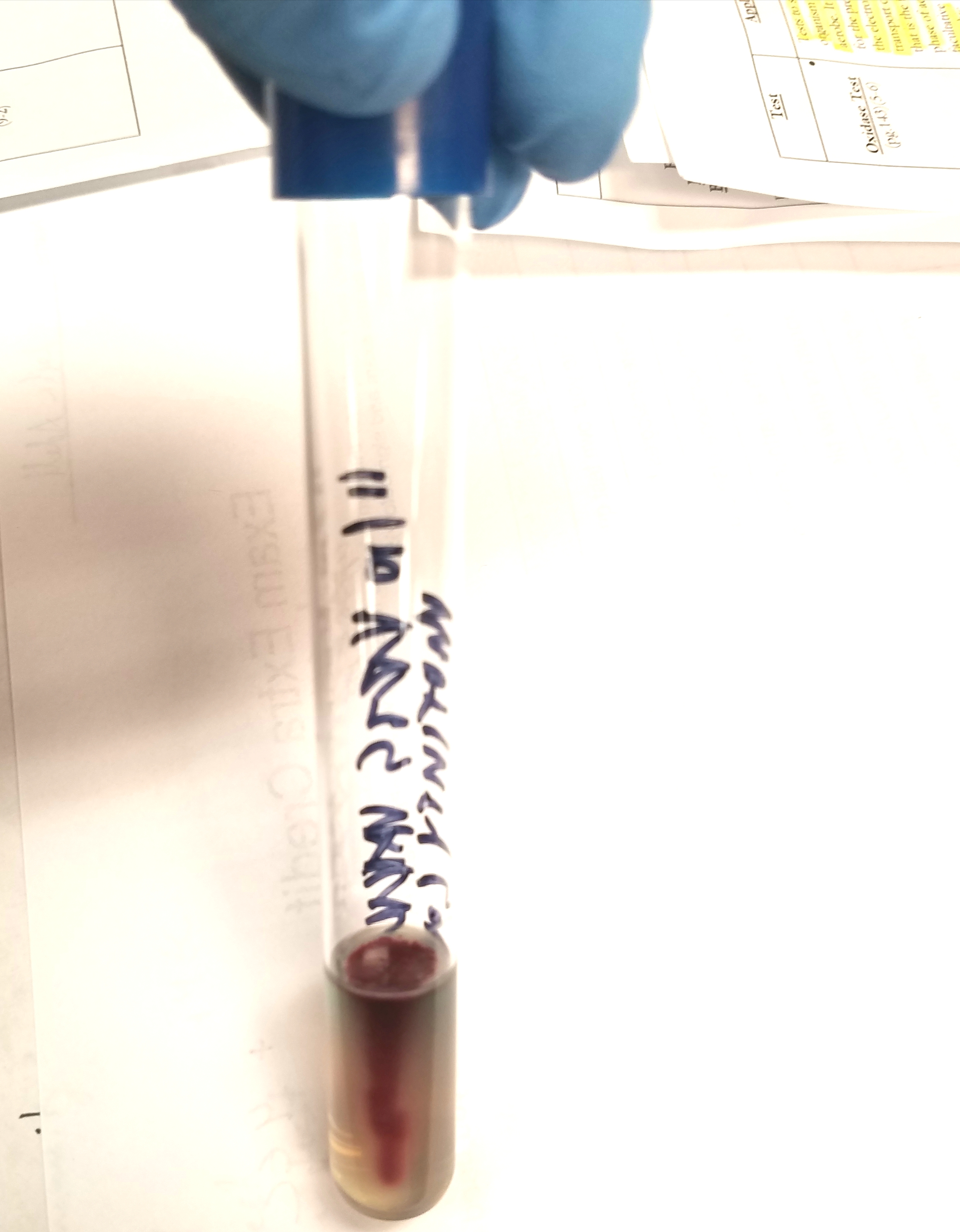
| 00:34, 7 December 2017 | Motilitypos.jpg (file) |  |
36 KB | Ladell | 1 | |
| 23:56, 6 December 2017 | 20171206 185213.jpg (file) |  |
1.38 MB | Ladell | 1 | |
| 23:35, 6 December 2017 | 20171206 183137.jpg (file) |  |
1.74 MB | Ladell | 1 | |
| 20:21, 6 December 2017 | Ecoli cell3.jpg (file) |  |
123 KB | Jljordan77 | 1 | |
| 20:18, 6 December 2017 | Ecoli cell2.jpg (file) |  |
8 KB | Jljordan77 | 1 | |
| 20:07, 6 December 2017 | Ecoli cell.jpg (file) |  |
38 KB | Jljordan77 | 1 | |
| 22:42, 5 December 2017 | Copperfield park photo.png (file) |  |
1.59 MB | Jljordan77 | 1 | |
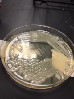
| 18:30, 5 December 2017 | Soil motilitySHWEB.jpg (file) |  |
1.98 MB | Marissa.parks | Positive motility results from soil sample, likely a Paenibacillus species. | 1 |
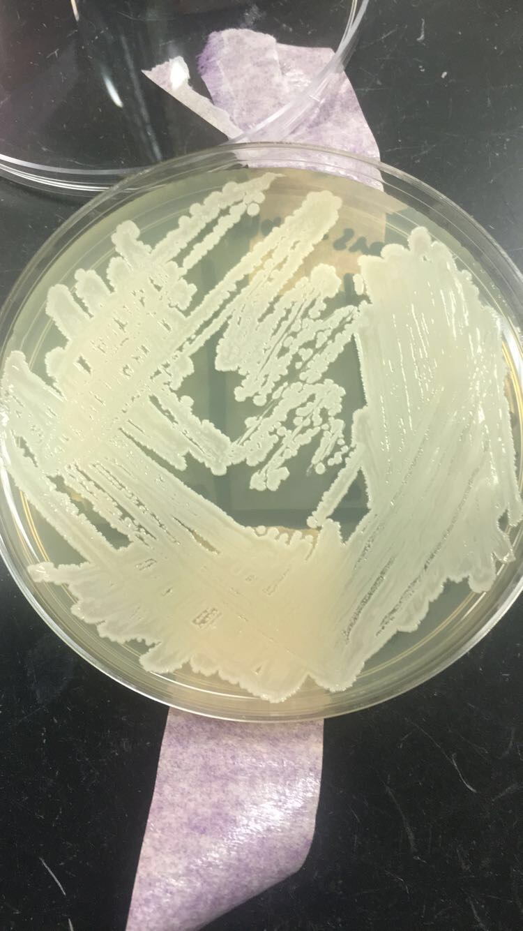
| 18:15, 5 December 2017 | Colony morphology soil SHWEB.jpg (file) |  |
1.95 MB | Marissa.parks | Paenibacillus colonies on LB agar. | 1 |
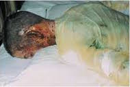
| 22:11, 3 December 2017 | P.aeruginosa burn victim.jpg (file) |  |
8 KB | Ladell | 1 | |
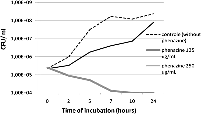
| 19:13, 1 December 2017 | Phenazine&MRSA.jpg (file) |  |
69 KB | Ladell | 1 | |
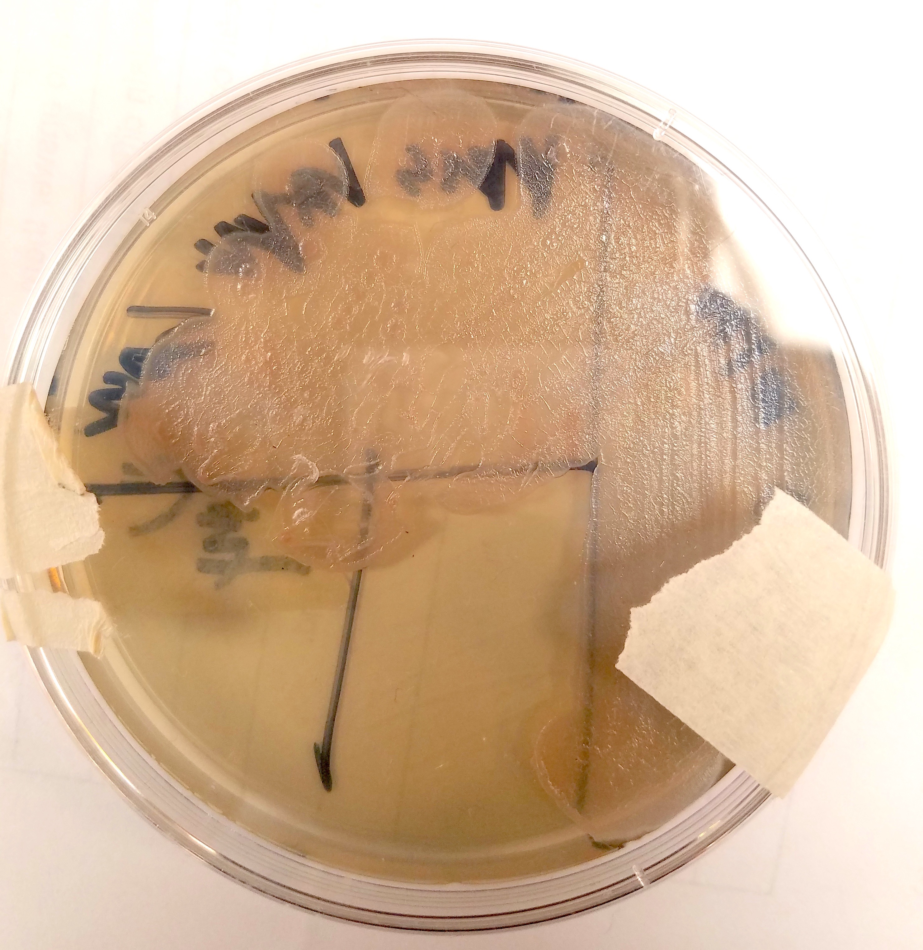
| 18:37, 1 December 2017 | P.aeruginosa37.jpg (file) |  |
1.78 MB | Ladell | P. aeruginosa incubated at 37 degrees celsius | 1 |
| 18:24, 1 December 2017 | P.aeruginosa30.jpg (file) |  |
1.89 MB | Ladell | P. aeruginosa incubated at 30 degrees celsius | 1 |
| 07:06, 28 November 2017 | IMG 3417.jpg (file) |  |
121 KB | Kbt17 | 1 | |
| 06:48, 28 November 2017 | IMG 3610.jpg (file) |  |
161 KB | Kbt17 | 1 | |
| 06:47, 28 November 2017 | IMG 3267.jpg (file) |  |
35 KB | Kbt17 | 1 | |
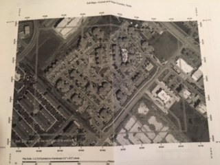
| 06:36, 28 November 2017 | SoilMap.jpg (file) |  |
37 KB | Kbt17 | 1 | |
| 06:28, 28 November 2017 | FullSizeRender.jpg (file) |  |
1.24 MB | Kbt17 | Soil Map Location | 1 |
| 05:53, 18 October 2017 | Pmicra.png (file) |  |
184 KB | S4359281 | 1 | |
| 05:50, 18 October 2017 | PMycra.png (file) |  |
336 KB | S4359281 | 1 | |
| 05:42, 18 October 2017 | Prevotella.jpg (file) |  |
22 KB | S4331513 | 1 | |
| 01:27, 15 October 2017 | Fusobacterium Periodonticum.png (file) |  |
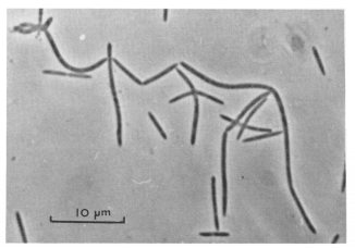
80 KB | S4471317 | Photomicrograph of “F. periodonticum” taken by Slots et.al <sup>[1]</sup>, the first finders of “F. periodonticum”. 1. [http://journals.sagepub.com/doi/abs/10.1177/00220345830620090901?url_ver=Z39.88-2003&rfr_id=ori:rid:crossre... | 1 |
