File list
From MicrobeWiki, the student-edited microbiology resource
This special page shows all uploaded files.
| Date | Name | Thumbnail | Size | User | Description | Versions |
|---|---|---|---|---|---|---|
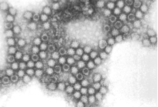
| 16:38, 6 December 2021 | FCV Macro Structure.jpg (file) |  |
56 KB | Joycez5 | 1 | |
| 16:36, 6 December 2021 | FCV Macrostructure.png (file) |  |
186 KB | Joycez5 | 1 | |
| 16:33, 6 December 2021 | Cat.jpg (file) |  |
40 KB | Joycez5 | 1 | |
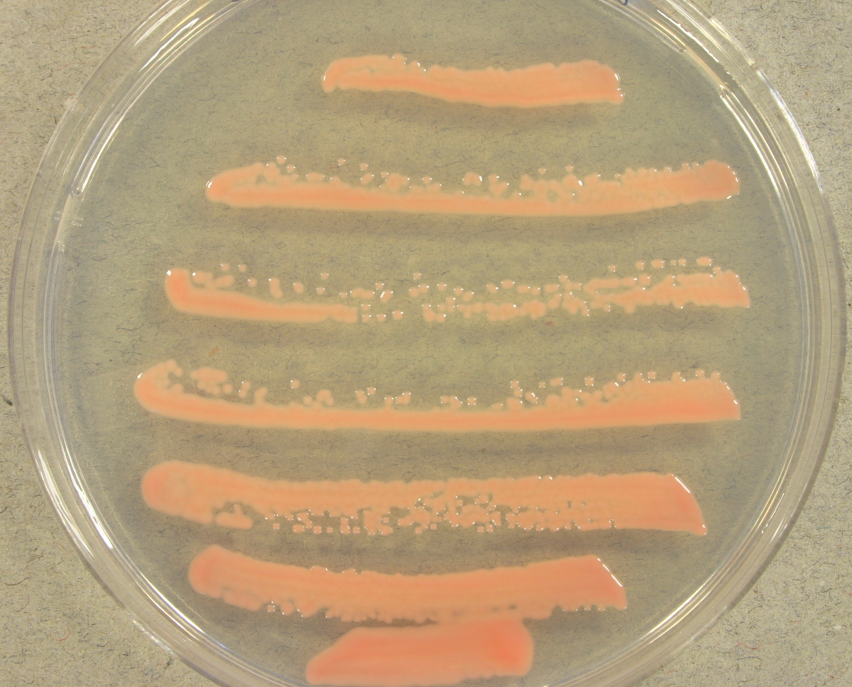
| 15:54, 6 December 2021 | Rhodotorula glutinis.jpeg (file) |  |
1.45 MB | Katchmar | Rhodotorula glutinis UAMH 2662, colony developed at 30 °C for 2 days on potato dextrose agar. | 1 |
| 15:36, 6 December 2021 | Goleva.png (file) |  |
1.41 MB | Moceyunas1 | 1 | |
| 15:24, 6 December 2021 | Akhabir.gif (file) |  |
37 KB | Moceyunas1 | 1 | |
| 14:51, 6 December 2021 | Pnas.2010761117fig01.png (file) |  |
75 KB | Ahysa | Figure 2. Phylogenomic analysis of several Mycena species and related fungi. Image created by Huei-Mien Ke, et al. Published November 23, 2020 by PNAS. Image licensed under CC BY-NC-ND 4.0. | 1 |
| 14:50, 6 December 2021 | 63ea46f0a1150c2d447e2e3796bc1a0e.jpg (file) |  |
76 KB | Campionm | 1 | |
| 14:46, 6 December 2021 | Shiitakemushrrom.jpg (file) |  |
32 KB | Nadifa | Figure 1 - Lentinula edodes (shiitake mushroom) morphology. Photo credit: International Press Telecommunications Council (IPTC) | 1 |
| 14:46, 6 December 2021 | JuninVirus.png (file) |  |
252 KB | Shahs07 | Figure 1: Visual representation of Junin Virus genome structure and life cycle. Adapted from Review of Mammarenavirus Biology and Replication[8] The virion first enters the host cell through endocytosis and then the RNA inside of it is released. There are two stages of mRNA: early and late. Early mRNA goes through NP and LP translation while late mRNA goes through Z translation. After translation, the proteins join together and the virion is assembled and released from the cell. Alongside thi... | 1 |
| 14:40, 6 December 2021 | Mycena chlorophos (Berk. & M.A. Curtis) Sacc 77081.jpg (file) | _Sacc_77081.jpg) |
282 KB | Ahysa | 1 | |
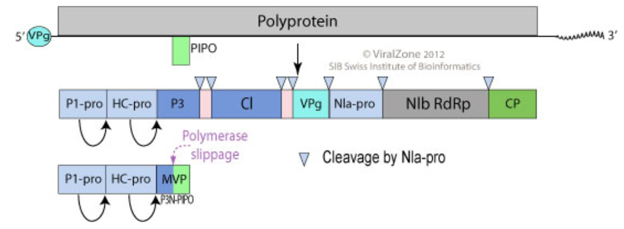
| 14:38, 6 December 2021 | PotyFigure1.png (file) |  |
236 KB | Szhang99 | 1 | |
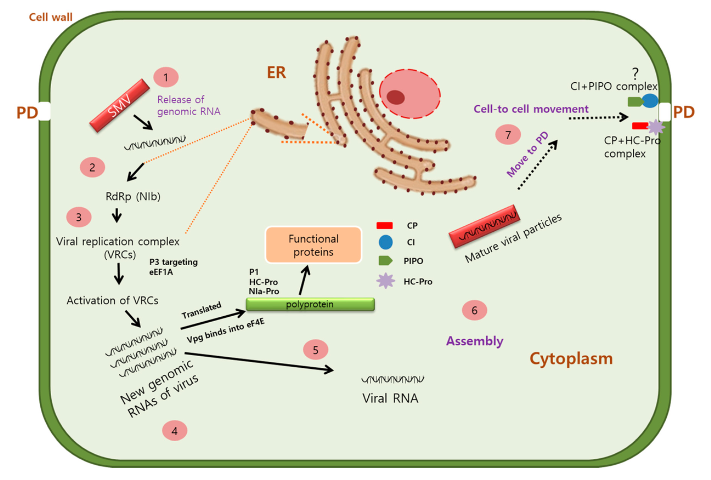
| 14:27, 6 December 2021 | PotyFigure2.png (file) |  |
663 KB | Szhang99 | 1 | |
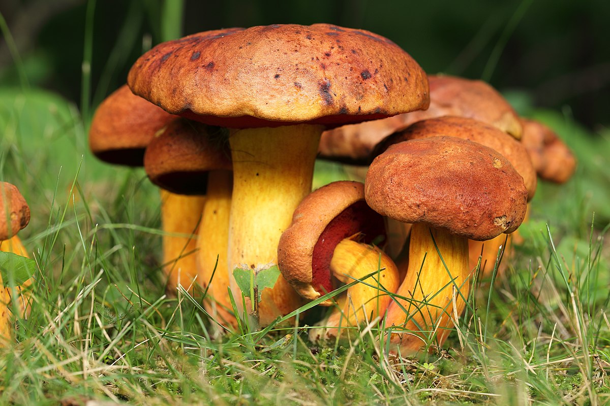
| 13:53, 6 December 2021 | Boletus spp UL 03.jpg (file) |  |
240 KB | Daly1 | 1 | |
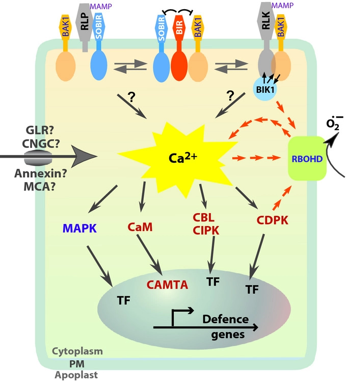
| 01:47, 6 December 2021 | PRR recognition to defence gene expression.webp (file) |  |
82 KB | Song3 | 1 | |
| 01:22, 6 December 2021 | PRRs recognize bacteria.jpg (file) |  |
56 KB | Song3 | 1 | |
| 01:15, 6 December 2021 | Ca2+-activated CaM binding to target domain.jpeg (file) |  |
1.19 MB | Song3 | 1 | |
| 15:52, 5 December 2021 | Fig2.png (file) |  |
15 KB | Ebudhram | 1 | |
| 15:43, 5 December 2021 | Fig1.png (file) |  |
175 KB | Ebudhram | 5 | |
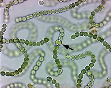
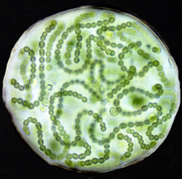
| 02:57, 5 December 2021 | Nostochetero.jpeg (file) |  |
20 KB | Tomasko1 | 1 | |
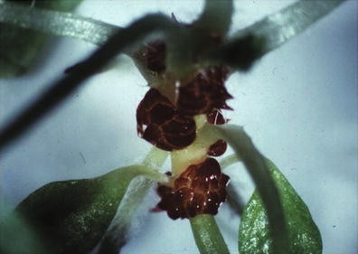
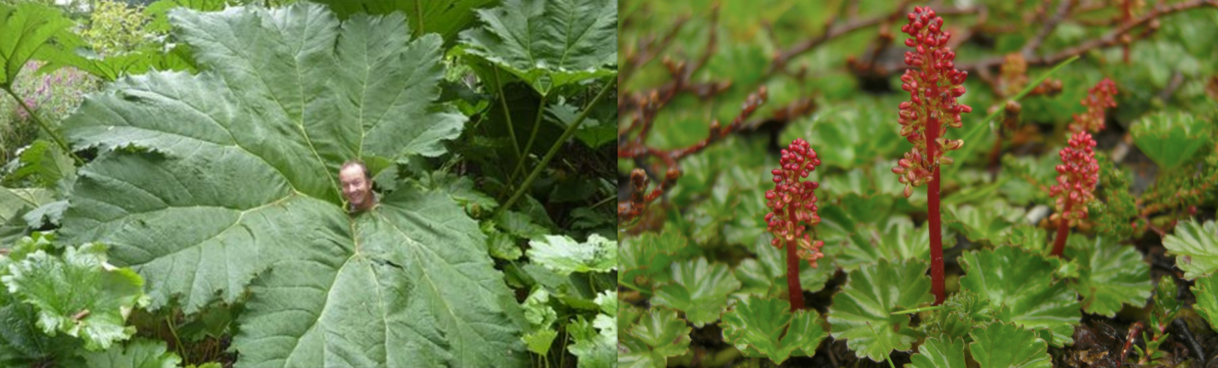
| 02:31, 5 December 2021 | Gunneraredgland.jpeg (file) |  |
19 KB | Tomasko1 | 1 | |
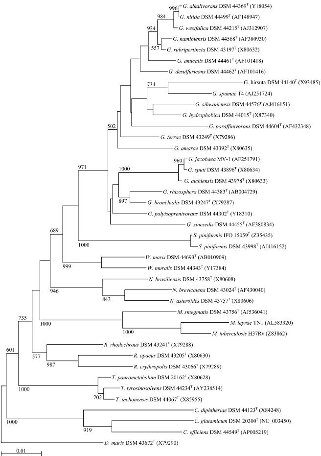
| 10:25, 3 December 2021 | Gordonia Phylogeny Tree.jpeg (file) |  |
76 KB | Xrodriguez2 | A phylogeny tree from figure 1 of the article https://www.ncbi.nlm.nih.gov/pmc/articles/PMC427784/ | 1 |
| 02:31, 2 December 2021 | 2,4-dinitrobenzenesulfonic acid.png (file) |  |
15 KB | Vanhorn1 | 1 | |
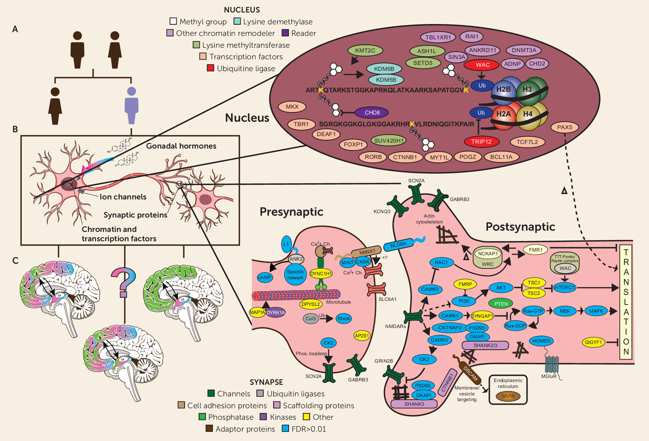
| 14:10, 1 December 2021 | Austism Genetics.jpeg (file) |  |
680 KB | Lowe1 | 1 | |
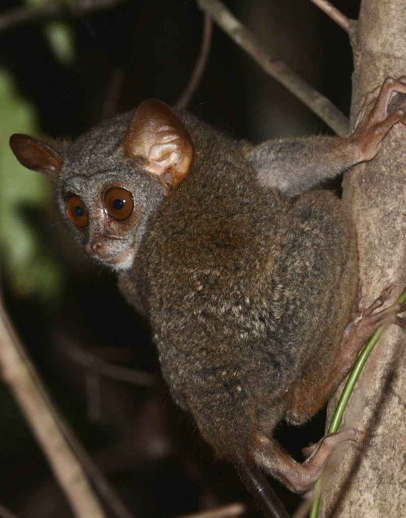
| 15:02, 29 November 2021 | Tarsierclingingbright.jpg (file) |  |
163 KB | Ford3 | 1 | |
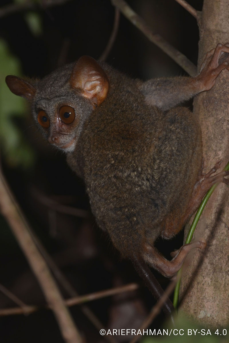
| 15:00, 29 November 2021 | Tarsierclinging.jpg (file) |  |
150 KB | Ford3 | 1 | |
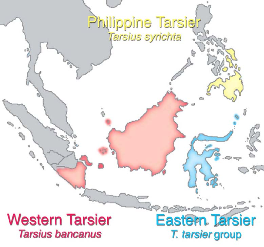
| 14:57, 29 November 2021 | Distribution-of-Extant-Tarsiers-The-northwestern-boundary-of-tarsiers-in-Sumatra-is.png (file) |  |
447 KB | Ford3 | 1 | |
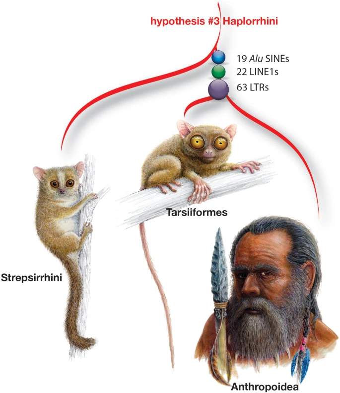
| 14:48, 29 November 2021 | Tarsierphylogeny.webp (file) |  |
76 KB | Ford3 | 1 | |
| 14:31, 29 November 2021 | Tarsier.jpg (file) |  |
188 KB | Ford3 | 1 | |
| 02:18, 29 November 2021 | Micrographs-of-Eggerthella-lenta-DSMZ-15644-cells-Ia-c-and-of-Slackia-equolifaciens.png (file) |  |
257 KB | Vanhorn1 | 1 | |
| 05:24, 27 November 2021 | Nostoc20.jpeg (file) |  |
69 KB | Tomasko1 | 1 | |
| 20:46, 26 November 2021 | Gunnera.png (file) |  |
1.89 MB | Tomasko1 | 1 | |
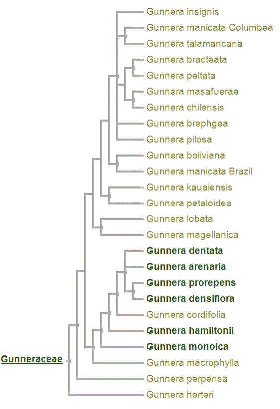
| 16:01, 26 November 2021 | Gunneratree.png (file) |  |
295 KB | Tomasko1 | 1 | |
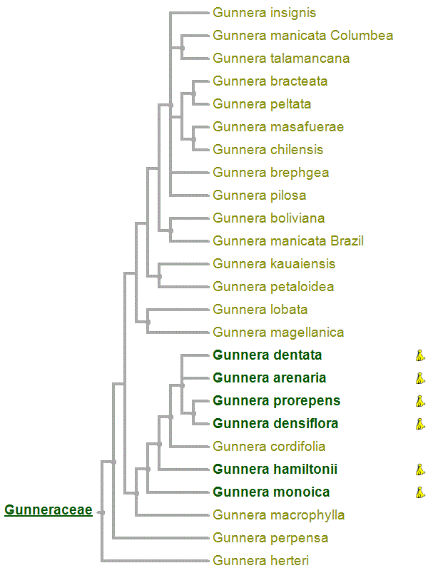
| 21:17, 24 November 2021 | ---Users-rachaeltomasko-Downloads-viewtree.gif (file) |  |
29 KB | Tomasko1 | 1 | |
| 21:52, 13 November 2021 | Prevotella22.jpg (file) |  |
12 KB | Vanhorn1 | 1 | |
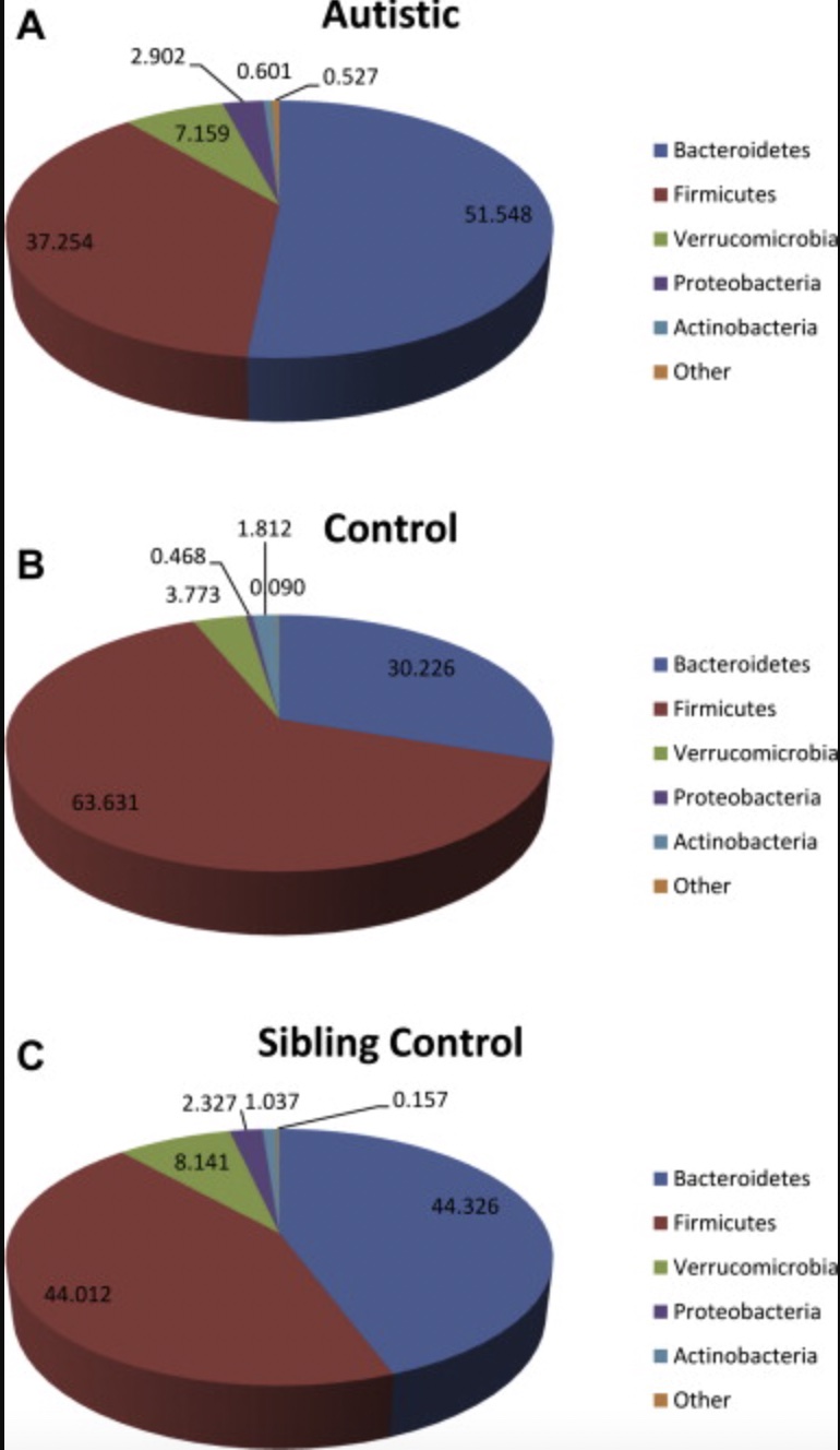
| 15:16, 11 November 2021 | JPiechart2.jpg (file) |  |
130 KB | Lowe1 | 1 | |
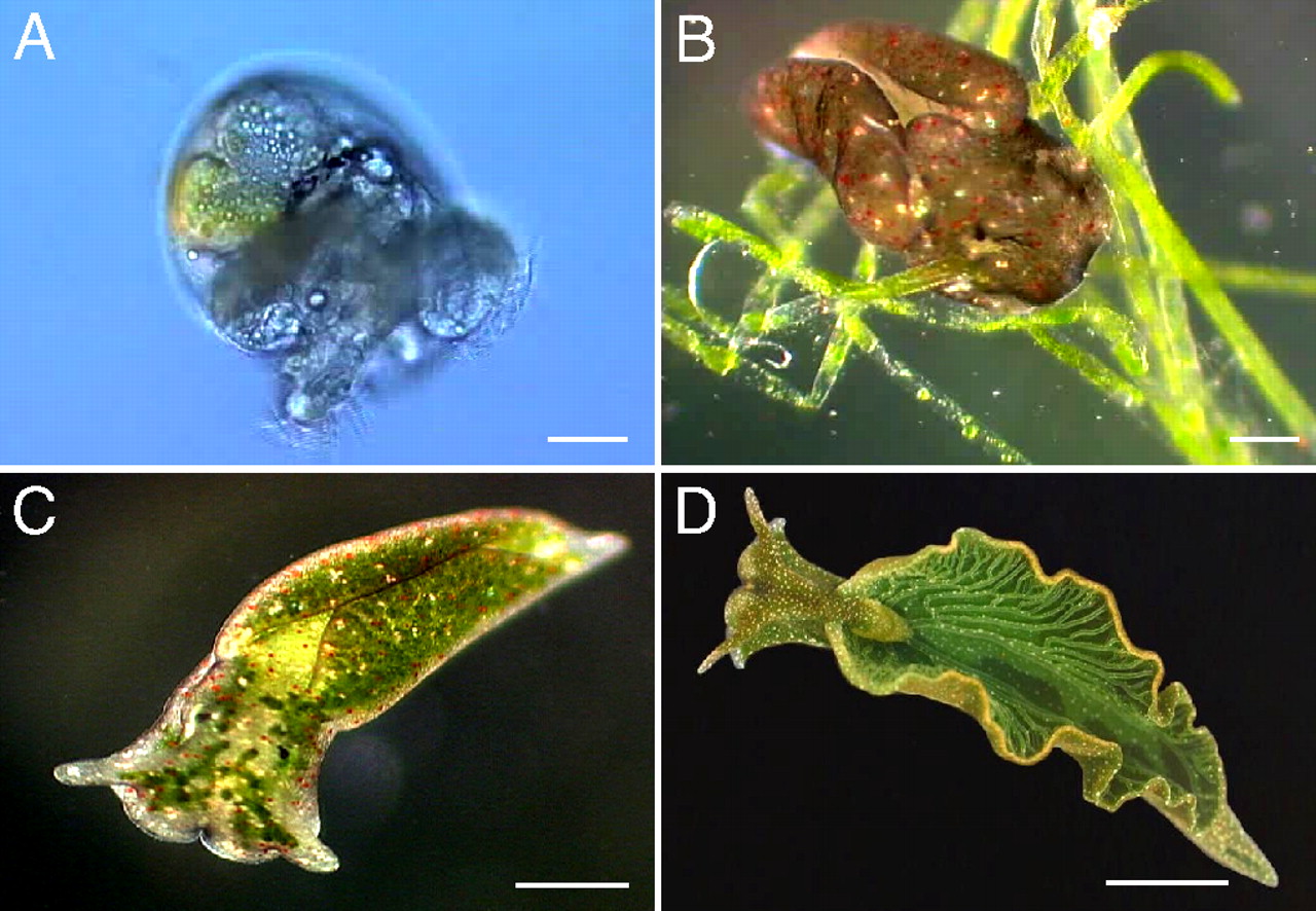
| 00:44, 8 November 2021 | Elysia chlorotica stages.jpg (file) |  |
258 KB | Yang4 | 1 | |
| 23:44, 7 November 2021 | Mfig001.jpeg (file) |  |
64 KB | Gusmano1 | Phenotypical traits expressed due to the homozygous inheritance of the successive nucleotide substitution c.453G>A and c.454C>A. | 1 |
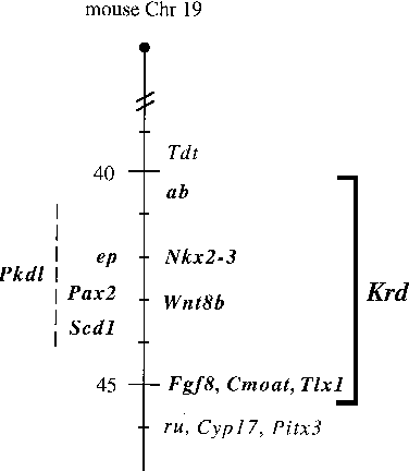
| 19:35, 7 November 2021 | Cr19.png (file) |  |
4 KB | Gusmano1 | General overview of top section of mouse Chromosome 19 including homeobox gene Pax2. | 1 |
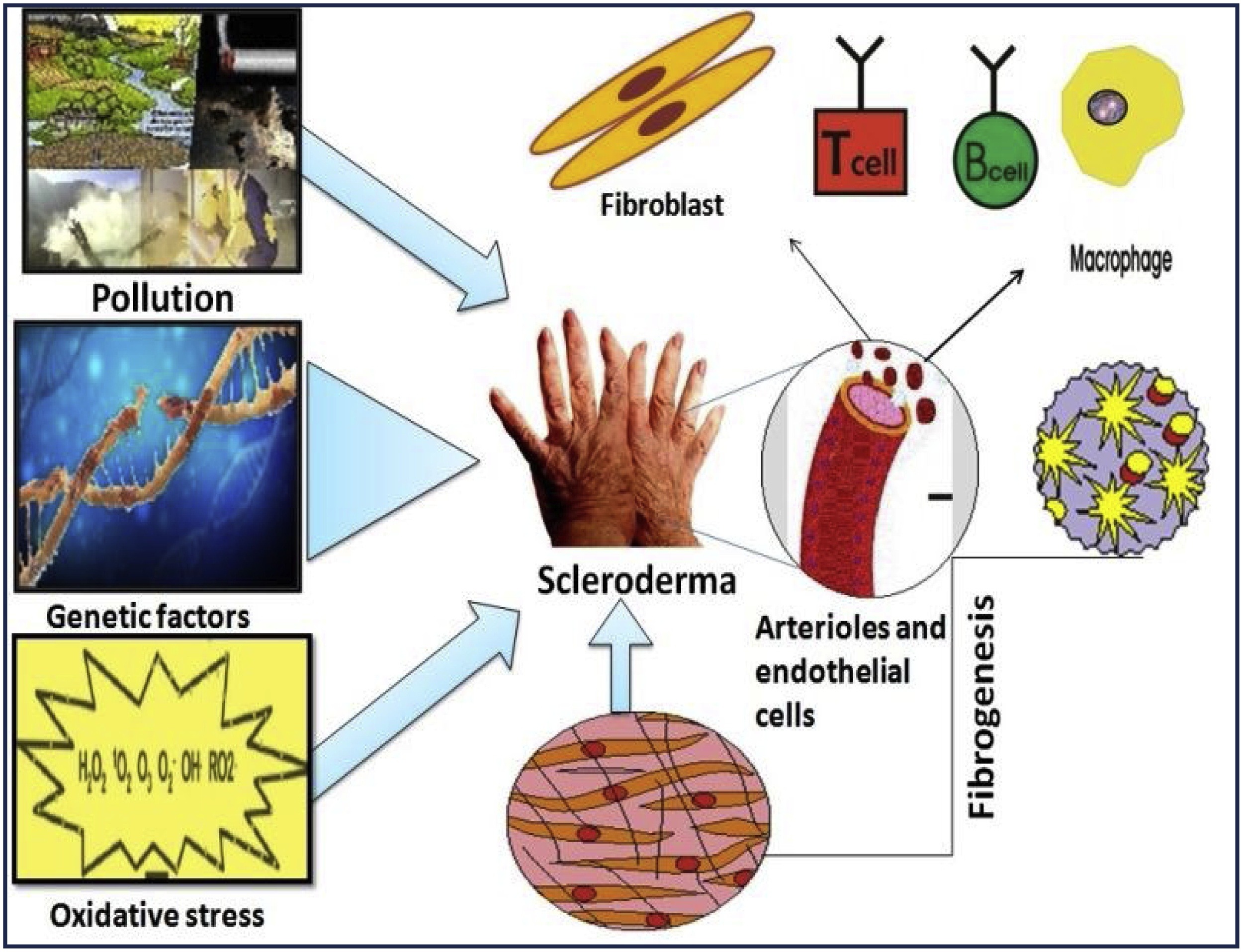
| 13:56, 7 November 2021 | Scleroderma Pathogenesis Image.jpg (file) |  |
741 KB | Vanhorn1 | 1 | |
| 17:24, 6 November 2021 | Https---animalia.us-east-1.linodeobjects.com-animals-photos-full-1.25x1-tarsier-tangkoko-national-park-sulawesi-indonesia.jpg (file) |  |
331 KB | Ford3 | 1 | |
| 19:24, 5 November 2021 | JPiechart.png (file) |  |
892 KB | Lowe1 | 1 | |
| 14:51, 5 November 2021 | Huffnagle-Dickson-Lukacs.jpg (file) |  |
37 KB | Moceyunas1 | 1 | |
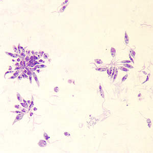
| 12:27, 5 November 2021 | Leishmania promastigote.jpg (file) |  |
11 KB | Pardue2 | 1 | |
| 12:23, 5 November 2021 | CaEntObPR22Gbv1akvPjtOOp8roGj8vMVwb098ov.jpeg (file) |  |
651 KB | Tomasko1 | 1 | |
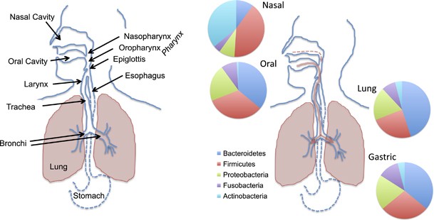
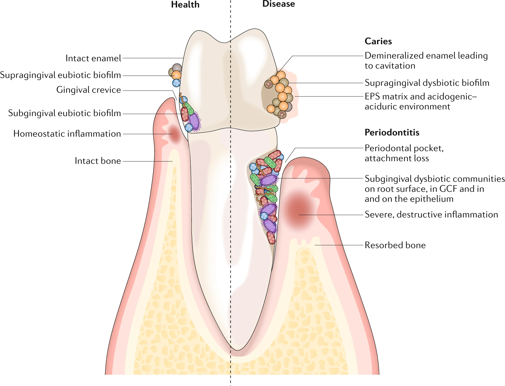
| 01:34, 31 August 2021 | Biogeography of oral microbiota.png (file) |  |
643 KB | A.reis | Figure 1: Microbial colonization occurs on all available surfaces, and microorganisms can also penetrate epithelial tissues and cells. The microbiota assembles into biofilm communities on the abiotic and biotic surfaces. In health (left), eubiotic biofilms maintain a homeostatic balance with the host. In disease (right), caries and periodontitis ensue when biofilms become dysbiotic, resulting in increased levels and duration of low pH challenge and the induction of destructive inflammatory re... | 1 |
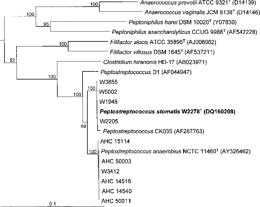
| 01:22, 31 August 2021 | Phylogenetic tree P stomatis.png (file) |  |
21 KB | A.reis | Fig. 1. Phylogenetic tree based on 16S rRNA gene sequence comparisons over 1283 aligned bases showing relationships between strains of P. anaerobius and P. stomatis sp. nov., Peptostreptococcus phylotype CK035 and related species. The tree was constructed using the neighbour-joining method following distance analysis of aligned sequences. Numbers represent bootstrap values for each branch based on data for 100 trees. Accession numbers for 16S rRNA gene sequences are given for each strain. Bar... | 1 |
| 20:33, 13 May 2021 | Darobactin Mode of Action.png (file) |  |
580 KB | Pintoned | Picture from: https://www.nature.com/articles/d41586-019-03730-x?draft=collection&fbclid=IwAR1G2xiGrA-d%20aXiitJe95nFJRSx5T3TiWWgsv5silORz9_E1PDET1n3uPxc | 1 |
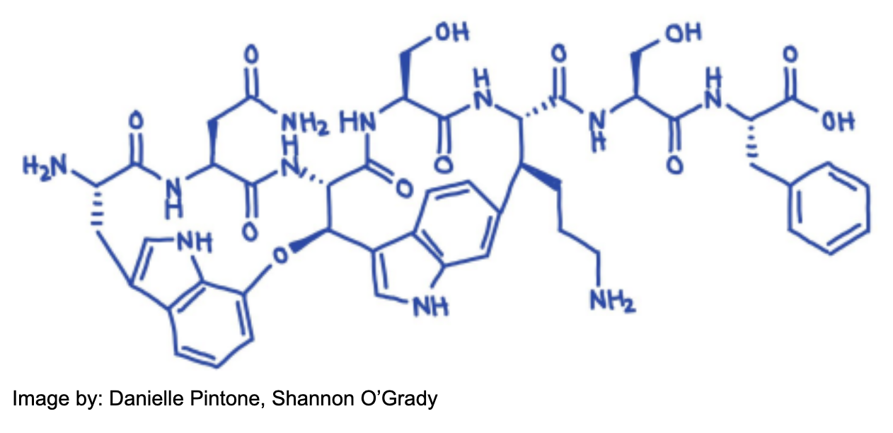
| 20:27, 13 May 2021 | Chemical Structure of Darobactin.png (file) |  |
346 KB | Pintoned | 1 | |
| 20:16, 13 May 2021 | Screen Shot 2021-05-13 at 4.11.29 PM.png (file) |  |
346 KB | Pintoned | 1 |
