File list
From MicrobeWiki, the student-edited microbiology resource
This special page shows all uploaded files.
| Date | Name | Thumbnail | Size | User | Description | Versions |
|---|---|---|---|---|---|---|
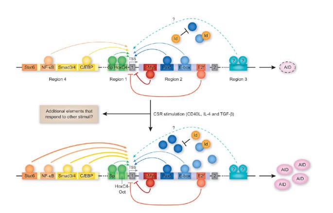
| 20:32, 9 December 2021 | AICDA gene depiction.jpg (file) |  |
40 KB | Smallwood1 | 1 | |
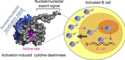
| 17:19, 9 December 2021 | AID deamination.jpg (file) |  |
69 KB | Smallwood1 | https://www.sciencedirect.com/science/article/pii/S0022283612008984 Activation Induced Cytidine Deaminase(AID) structure and mechanism | 1 |
| 03:44, 9 December 2021 | Deltaretrovirus virion.jpg (file) |  |
74 KB | Srnka1 | 1 | |
| 02:49, 9 December 2021 | 978-9283201342-C012-F001.002.jpg (file) |  |
71 KB | Srnka1 | 1 | |
| 01:25, 9 December 2021 | LuXOpLL21.png (file) |  |
302 KB | Lehr1 | 1 | |
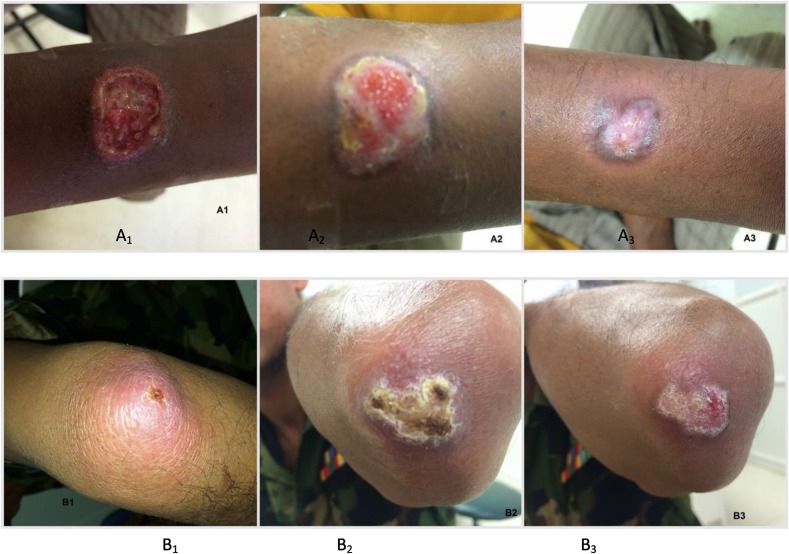
| 01:02, 9 December 2021 | Lesions.jpg (file) |  |
119 KB | Pardue2 | 1 | |
| 00:07, 9 December 2021 | Pm.jpg (file) |  |
9 KB | Blackwellscott1 | 1 | |
| 00:02, 9 December 2021 | Catbacteria.jpg (file) |  |
15 KB | Blackwellscott1 | 1 | |
| 22:28, 8 December 2021 | Elysia starvation.png (file) |  |
909 KB | Yang4 | 1 | |
| 19:35, 8 December 2021 | Unnamed.jpg (file) |  |
39 KB | Blackwellscott1 | cat breed | 1 |
| 19:33, 8 December 2021 | Fake cats.jpg (file) |  |
324 KB | Blackwellscott1 | 1 | |
| 19:29, 8 December 2021 | Images.jpg (file) |  |
9 KB | Blackwellscott1 | 2 | |
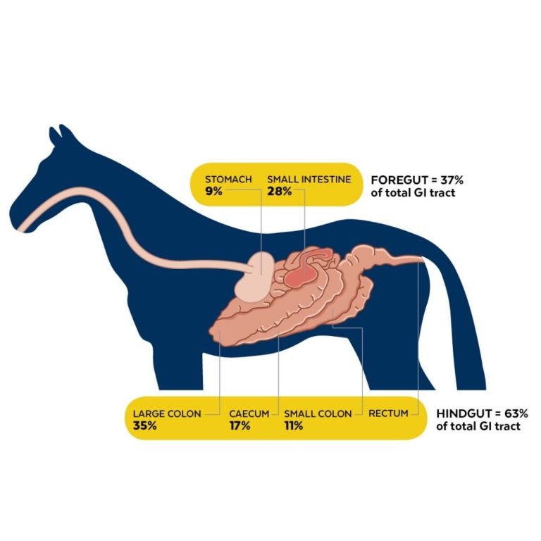
| 18:47, 8 December 2021 | EquineGut.jpg (file) |  |
43 KB | Childs1 | 1 | |
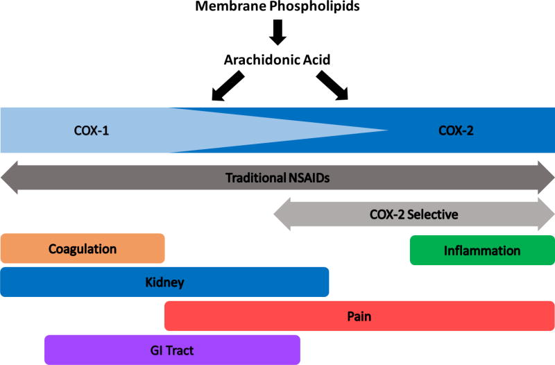
| 17:49, 8 December 2021 | COX.jpg (file) |  |
44 KB | Childs1 | 1 | |
| 14:22, 8 December 2021 | LuciferaseLL21.png (file) |  |
258 KB | Lehr1 | 1 | |
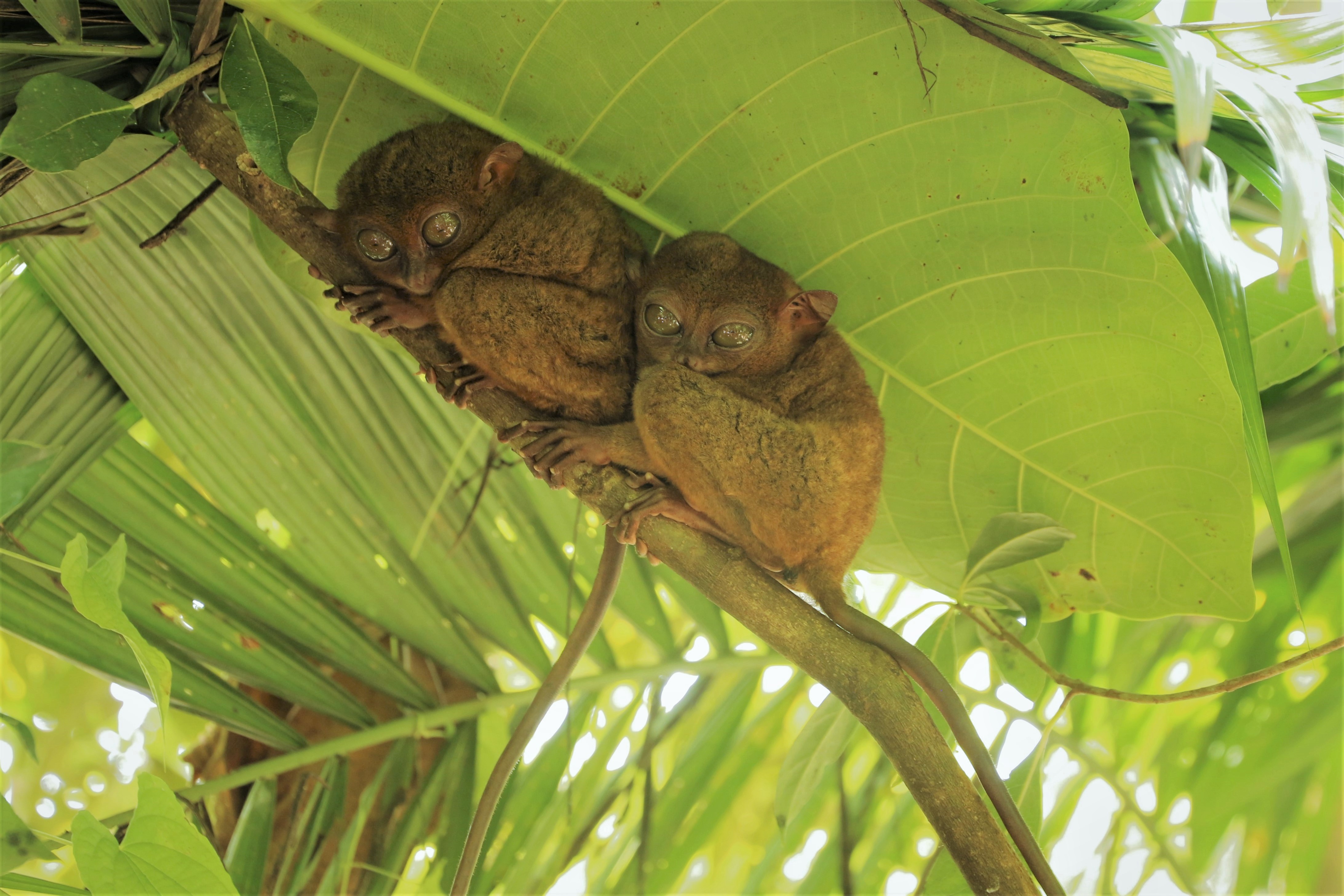
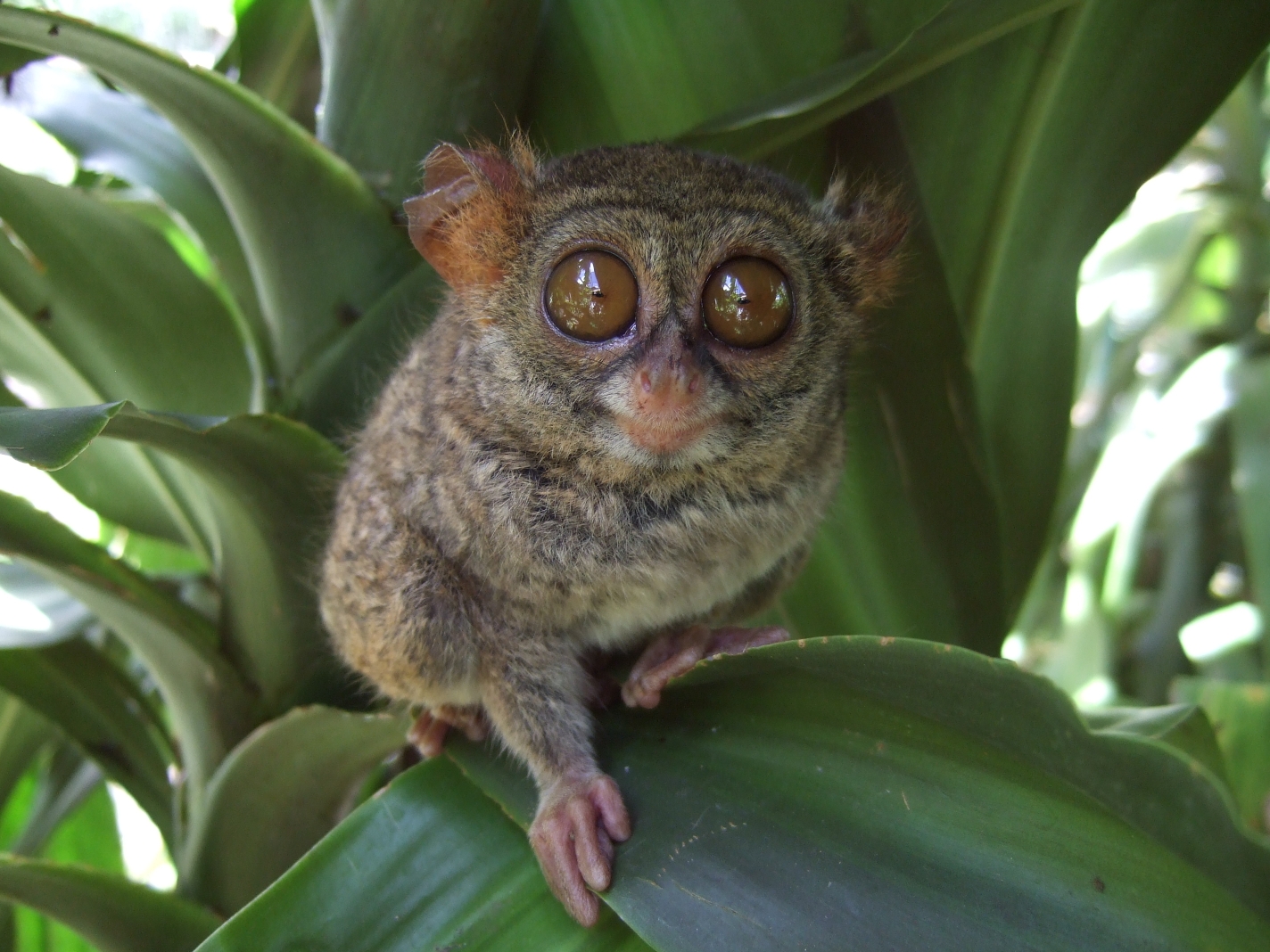
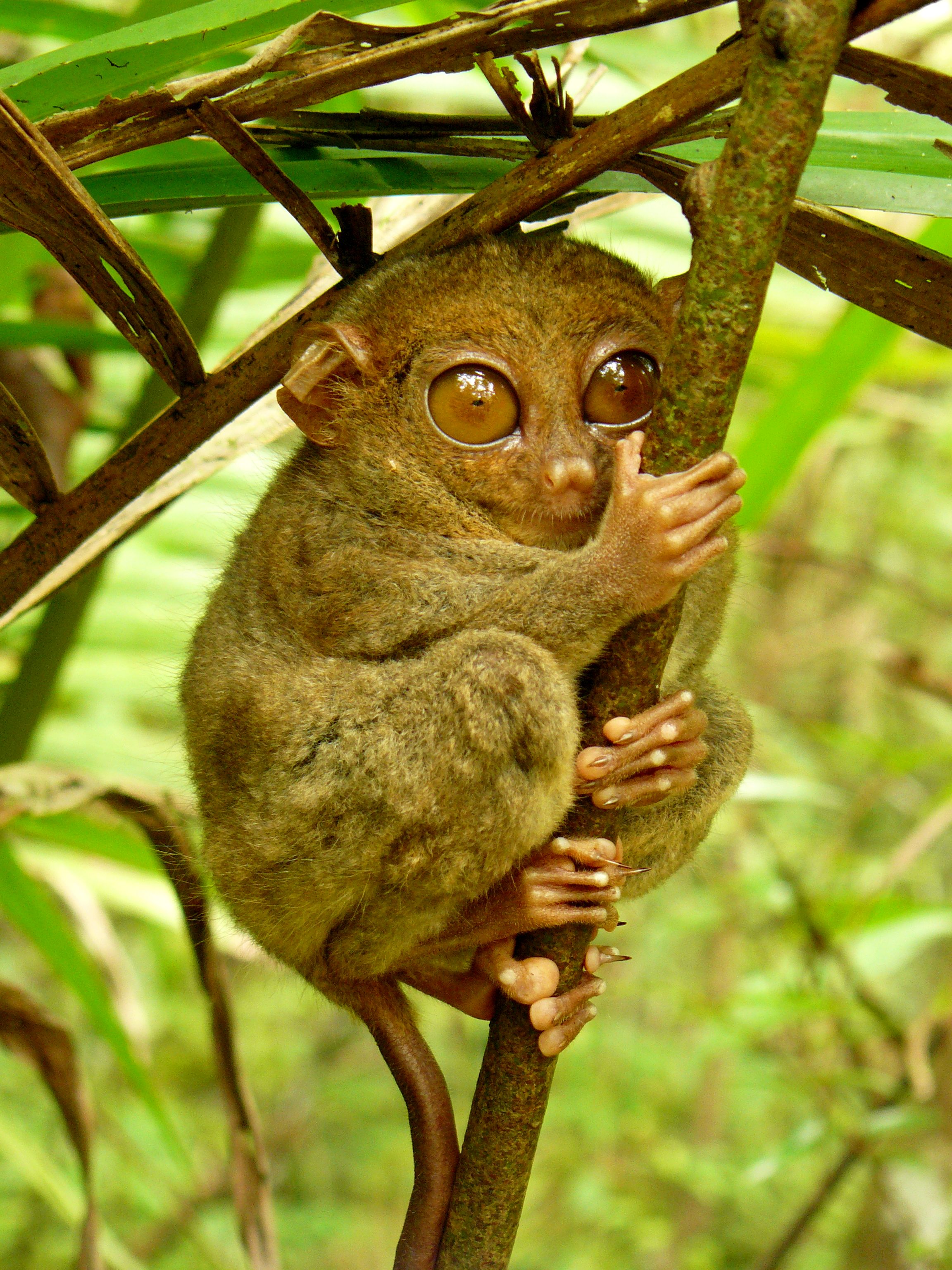
| 13:54, 8 December 2021 | Tarsiercouple.jpeg (file) |  |
1.45 MB | Ford3 | 1 | |
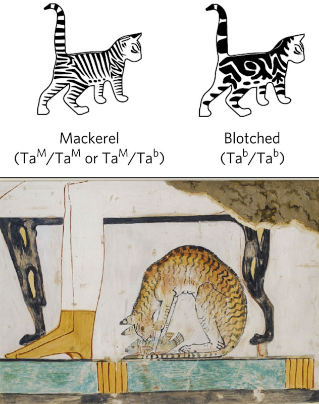
| 13:48, 8 December 2021 | Development-of-the-blotched-tabby-domestic-cat.jpg (file) |  |
117 KB | Blackwellscott1 | 1 | |
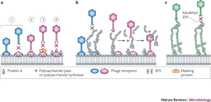
| 13:45, 8 December 2021 | Phage resistance.jpeg (file) |  |
48 KB | Somadder1 | 1 | |
| 13:42, 8 December 2021 | Tarsierleaf.jpg (file) |  |
993 KB | Ford3 | 1 | |
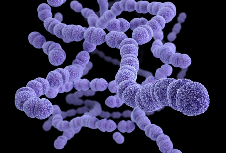
| 13:32, 8 December 2021 | Pneumococcal-v1-1.jpeg (file) |  |
103 KB | Somadder1 | 1 | |
| 13:30, 8 December 2021 | QSluxOp.jpg (file) |  |
40 KB | Lehr1 | 1 | |
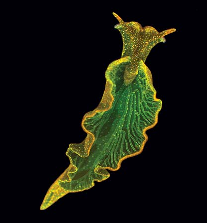
| 13:28, 8 December 2021 | Elysia chlorotica.jpeg (file) |  |
16 KB | Yang4 | 1 | |
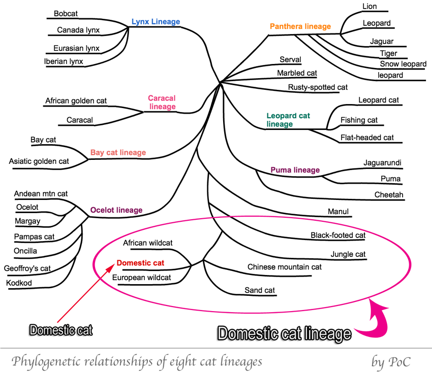
| 13:27, 8 December 2021 | Domestic.Cat.12.png (file) |  |
173 KB | Blackwellscott1 | Cat Phylogeny from Cat Chit Chat. | 1 |
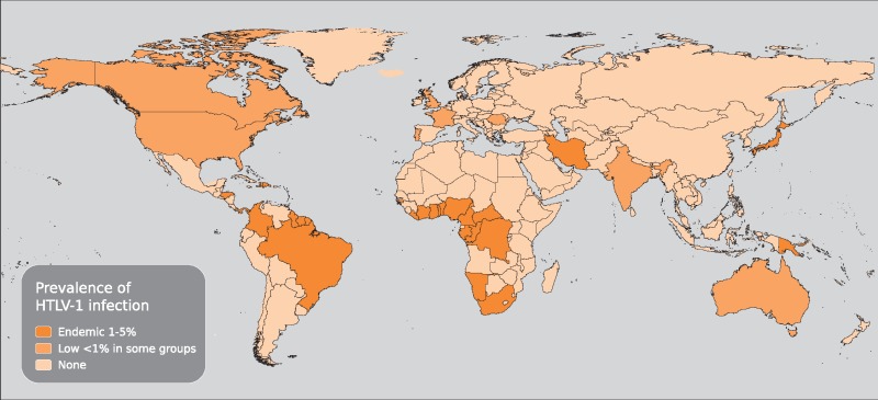
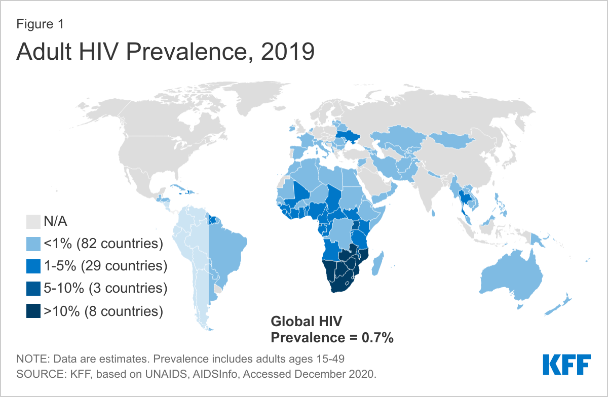
| 13:26, 8 December 2021 | Adult-hiv-prevalence-2019.png (file) |  |
205 KB | Timken1 | 1 | |
| 13:26, 8 December 2021 | Viruses- 2.jpg (file) |  |
217 KB | Srnka1 | 1 | |
| 13:25, 8 December 2021 | Vax1Regulation.png (file) |  |
1.4 MB | Gusmano1 | 2 | |
| 13:23, 8 December 2021 | Tarsier Hugs Mossy Branch.jpg (file) |  |
1.15 MB | Ford3 | 1 | |
| 13:22, 8 December 2021 | KaranWikiImage.png (file) |  |
14 KB | Mehta1 | 1 | |
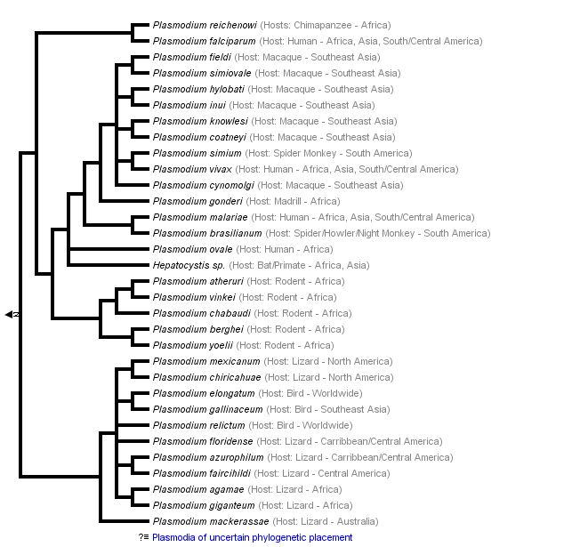
| 13:17, 8 December 2021 | Plasmodium.png (file) |  |
14 KB | Mehta1 | 2 | |
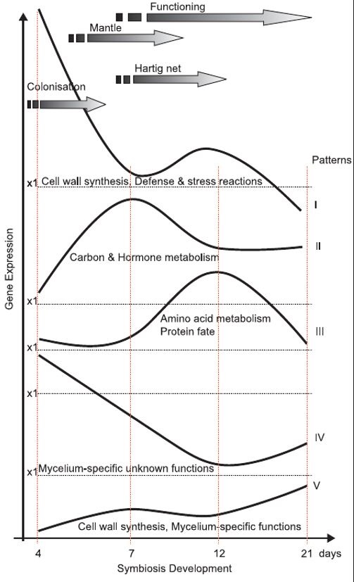
| 06:20, 8 December 2021 | GeneExprDev.JPG (file) |  |
65 KB | Daly1 | 1 | |
| 03:12, 8 December 2021 | SQUID.jpg (file) |  |
542 KB | Lehr1 | 1 | |
| 18:10, 7 December 2021 | BordeauxMixture.png (file) |  |
1.7 MB | Liu11 | 1 | |
| 18:09, 7 December 2021 | C.vulgaris.png (file) |  |
1.19 MB | Liu11 | 2 | |
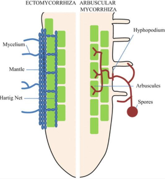
| 05:04, 7 December 2021 | HartigNet.JPG (file) |  |
38 KB | Daly1 | 1 | |
| 22:03, 6 December 2021 | Basidium schematic.svg.png (file) |  |
263 KB | Ahysa | 1 | |
| 21:58, 6 December 2021 | Gene E M Chloro.png (file) |  |
260 KB | Ahysa | 1 | |
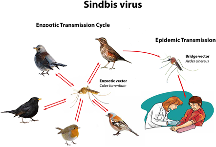
| 18:25, 6 December 2021 | Visual diagram.png (file) |  |
84 KB | Vnielsen | Figure 3 [Image reprinted under Creative Commons License (21)] Visual diagram of the primary route for Sindbis virus infection of humans through asymptomatic mammalian and insect carriers. Different species of birds and mosquitoes transmit the virus to each other. Mosquitoes infect humans, but humans cannot transmit Sindbis virus back to mosquitoes because of SINV’s naturally low viral load. | 1 |
| 18:09, 6 December 2021 | Billede1.jpg (file) |  |
14 KB | Vnielsen | Figure 2 [Image reprinted under Creative Commons License (13)]. Cryo-electron microscopy of the Sindbis virus - liposome complex. | 1 |
| 17:59, 6 December 2021 | FCV.jpeg (file) |  |
64 KB | Hkim512 | 1 | |
| 16:50, 6 December 2021 | Fcv.png (file) |  |
353 KB | Joycezhu | Figure 1: An electron micrograph revealing the macrostructure of FCV. The virion is typically 35-40nm in diameter with cup-like surface depressions (CDC). | 1 |
| 16:38, 6 December 2021 | FCV Macro Structure.jpg (file) |  |
56 KB | Joycez5 | 1 | |
| 16:36, 6 December 2021 | FCV Macrostructure.png (file) |  |
186 KB | Joycez5 | 1 | |
| 16:33, 6 December 2021 | Cat.jpg (file) |  |
40 KB | Joycez5 | 1 | |
| 15:54, 6 December 2021 | Rhodotorula glutinis.jpeg (file) |  |
1.45 MB | Katchmar | Rhodotorula glutinis UAMH 2662, colony developed at 30 °C for 2 days on potato dextrose agar. | 1 |
| 15:36, 6 December 2021 | Goleva.png (file) |  |
1.41 MB | Moceyunas1 | 1 | |
| 15:24, 6 December 2021 | Akhabir.gif (file) |  |
37 KB | Moceyunas1 | 1 | |
| 14:51, 6 December 2021 | Pnas.2010761117fig01.png (file) |  |
75 KB | Ahysa | Figure 2. Phylogenomic analysis of several Mycena species and related fungi. Image created by Huei-Mien Ke, et al. Published November 23, 2020 by PNAS. Image licensed under CC BY-NC-ND 4.0. | 1 |
| 14:50, 6 December 2021 | 63ea46f0a1150c2d447e2e3796bc1a0e.jpg (file) |  |
76 KB | Campionm | 1 | |
| 14:46, 6 December 2021 | Shiitakemushrrom.jpg (file) |  |
32 KB | Nadifa | Figure 1 - Lentinula edodes (shiitake mushroom) morphology. Photo credit: International Press Telecommunications Council (IPTC) | 1 |
| 14:46, 6 December 2021 | JuninVirus.png (file) |  |
252 KB | Shahs07 | Figure 1: Visual representation of Junin Virus genome structure and life cycle. Adapted from Review of Mammarenavirus Biology and Replication[8] The virion first enters the host cell through endocytosis and then the RNA inside of it is released. There are two stages of mRNA: early and late. Early mRNA goes through NP and LP translation while late mRNA goes through Z translation. After translation, the proteins join together and the virion is assembled and released from the cell. Alongside thi... | 1 |
