File list
From MicrobeWiki, the student-edited microbiology resource
This special page shows all uploaded files.
| Date | Name | Thumbnail | Size | User | Description | Versions |
|---|---|---|---|---|---|---|
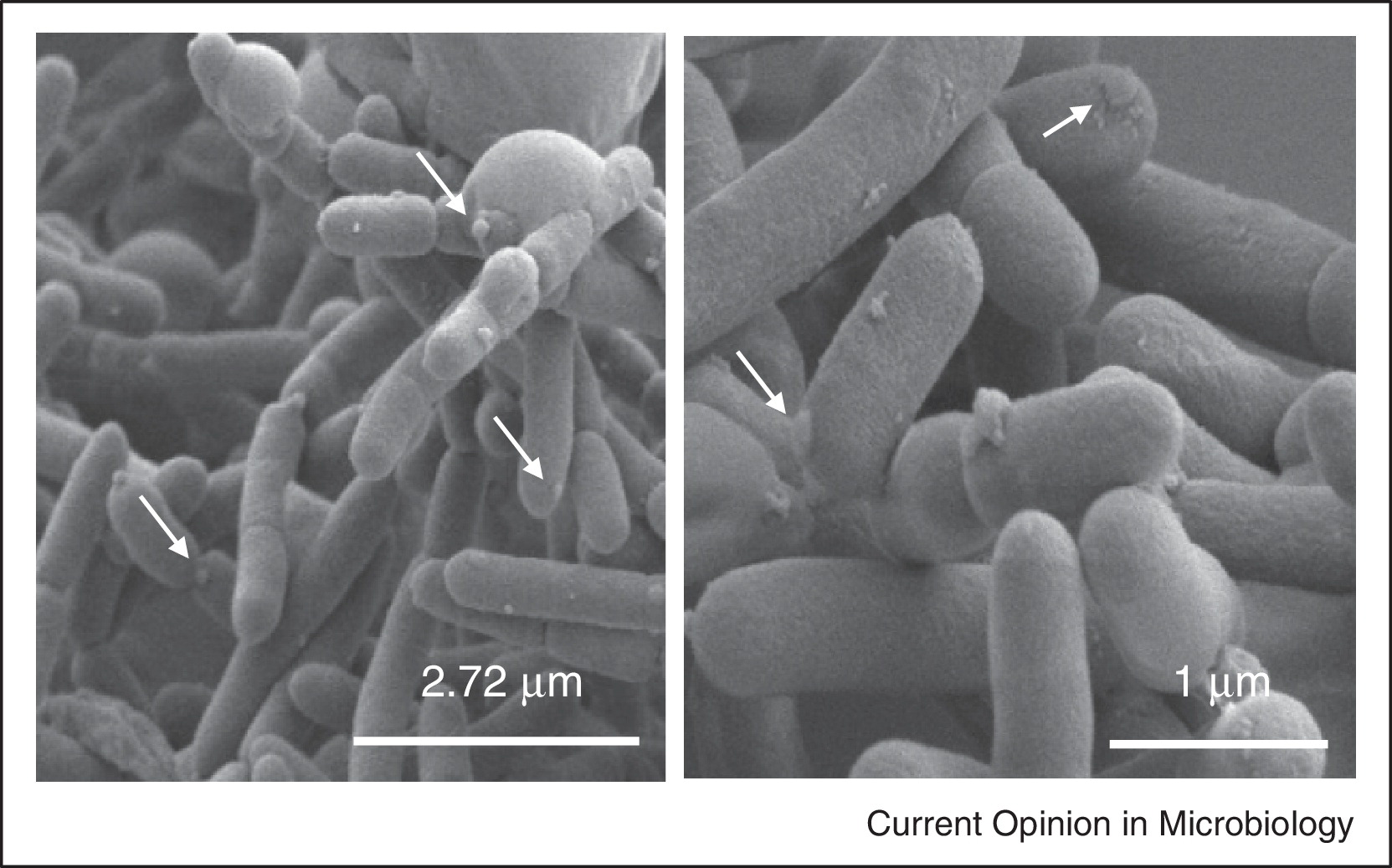
| 15:54, 23 February 2024 | 1-s2.0-S1369527413000775-gr1 lrg.jpg (file) |  |
294 KB | Arone1 | 1 | |
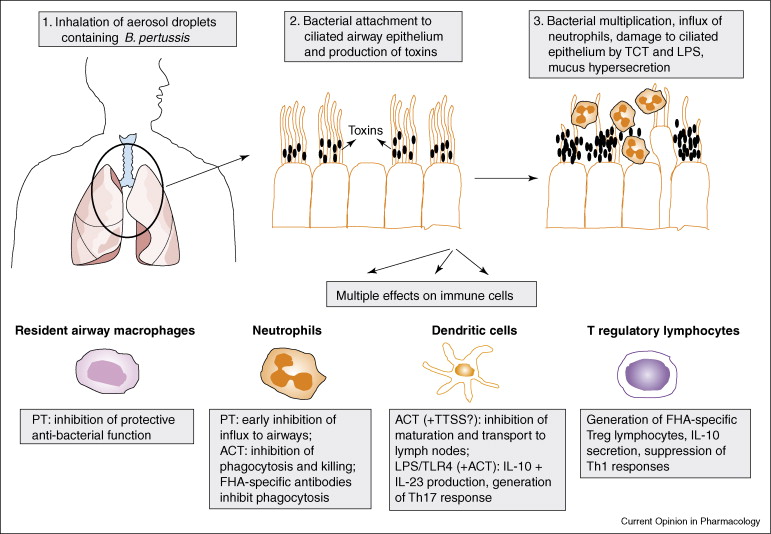
| 18:47, 14 April 2022 | 1-s2.0-S1471489207000598-gr1.jpg (file) |  |
95 KB | White4 | 1 | |
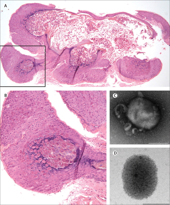
| 00:47, 26 April 2016 | 1-s2.0-S1473309913701099-gr2.jpg (file) |  |
195 KB | Mairsone | 1 | |
| 00:43, 26 April 2016 | 1-s2.0-S1473309913701099-gr3.jpg (file) |  |
350 KB | Mairsone | 1 | |
| 07:54, 12 December 2012 | 1-s2.0-S153561080400337X-gr1.jpg (file) |  |
36 KB | OconnorMC | 1 | |
| 21:41, 12 December 2022 | 1-s2.0-S2214250918300775-gr1-2.jpg (file) |  |
25 KB | Mjm2111 | 1 | |
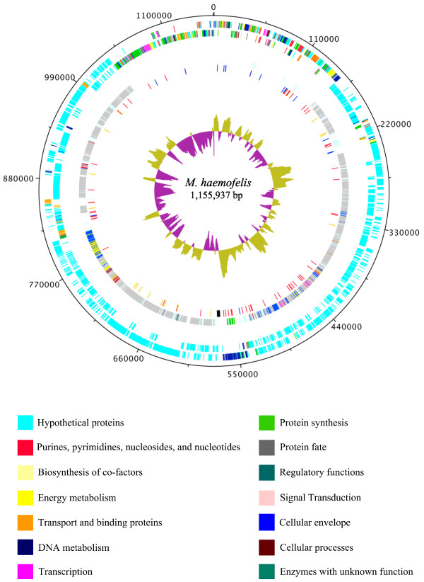
| 19:07, 5 December 2022 | 1-s2.0-S2214662820300426-gr1 lrg.jpg (file) |  |
547 KB | Liu13 | 1 | |
| 05:49, 6 December 2019 | 1-s2.0-S2468233017300634-gr4.jpg (file) |  |
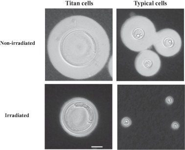
19 KB | Neal2 | Titan cells of C. neoformans shown next to typical C. neoformans cells | 1 |
| 03:04, 14 December 2012 | 1.2SME media.png (file) |  |
9 KB | Dominiquehaag | 1 | |
| 06:58, 24 May 2020 | 1. Dallol hydrothermal fields (Kotopoulou et al. 2018).jpeg (file) | .jpeg) |
392 KB | Rachelso2020 | The Dallol hydrothermal system is a high temperature, hypersaline, and hyperacidic environment located in the Danakil Depression in northeastern Ethiopia. Source: https://pubs.acs.org/doi/10.1021/acsearthspacechem.8b00141 | 1 |
| 23:34, 21 November 2013 | 1.jpg (file) |  |
45 KB | Salmamashkoor1 | 2 | |
| 00:03, 11 December 2018 | 1.png (file) |  |
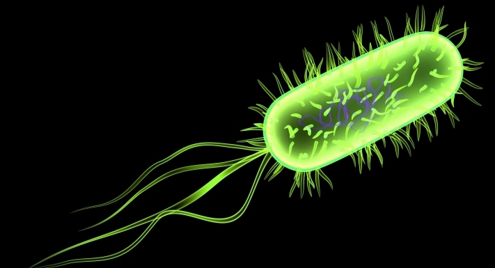
1.05 MB | Ij1212 | Image 1. The image above represents basic morphology of Campylobacter; they are gram negative(pink), spiral shaped rods (13). | 2 |
| 03:54, 2 May 2013 | 1.pptx.png (file) |  |
133 KB | Safa.alqayyar | 1 | |
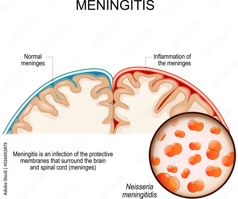
| 00:45, 14 April 2024 | 1000 F 526052873 t4gWHCRTR3vOE1UPwumyx9LDJZbMfJs1.jpg (file) |  |
218 KB | Gest1 | 1 | |
| 18:43, 31 May 2006 | 1002L.jpg (file) |  |
12 KB | Tashiror | 1 | |
| 01:14, 29 August 2008 | 10043 lores.jpg (file) |  |
63 KB | Jil014 | 1 | |
| 10:59, 16 July 2013 | 1006listeria.jpg (file) |  |
88 KB | Jennifer.E.Gallup-1 | 1 | |
| 17:35, 26 March 2013 | 100907a.jpg (file) |  |
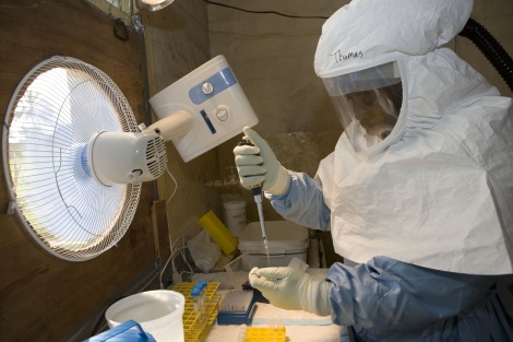
64 KB | Vgawlik6782 | Photo: Globalhealth.gov | 1 |
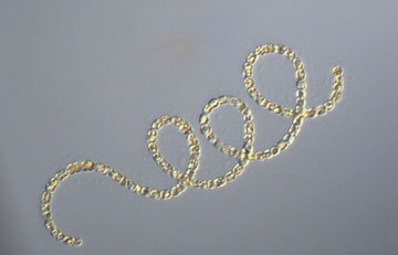
| 16:56, 16 April 2023 | 10179 lores.jpg (file) |  |
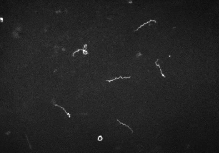
31 KB | Cenizalevine1 | Treponema pallidum dark field microscopy from the CDC, circa 1961. | 1 |
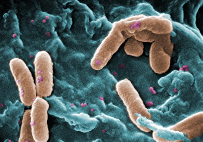
| 08:07, 28 August 2009 | 10247 lores.jpg (file) |  |
90 KB | Mhigashi | This scanning electron micrograph (SEM) depicted three views of a single Gram-negative Neisseria gonorrhoeae bacterium. Under this highly-magnified view, the “roughened” texture of the bacterium’s cell wall is made visible. As a Gram-negative bacter | 1 |
| 14:08, 29 April 2014 | 10306957 10152185468794475 448335891 a.jpg (file) |  |
4 KB | Maasstev | Methanogenium Frigidum | 1 |
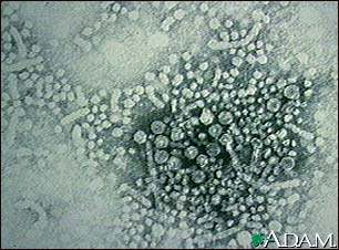
| 14:55, 6 June 2006 | 1031.jpg (file) |  |
20 KB | Mschlemm | Hepatitis B virus. From [http://www.nlm.nih.gov/medlineplus/ency/imagepages/1031.htm Medline Plus] | 1 |
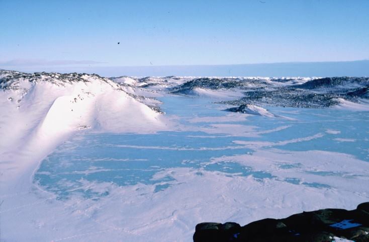
| 14:00, 29 April 2014 | 10323022 10152185468789475 949939896 n.jpg (file) |  |
39 KB | Maasstev | Ace Lake, Antarctuca | 1 |
| 21:25, 6 December 2008 | 1037 lores.jpg (file) |  |
12 KB | Considinek | 1 | |
| 16:18, 8 November 2020 | 10815 lores.jpg (file) |  |
59 KB | Slonczewski | CDC by Frederick A. Murphy. Colorized transmission electron microscopic image showing the filamentous and curved morphology of an Ebola virus particle. See PHIL 1181 for a black and white version of this image. | 1 |
| 20:59, 18 April 2018 | 108ef8b8771ca41a65d7acc195ab25f9.jpg (file) |  |
281 KB | Jesszell1 | 1 | |
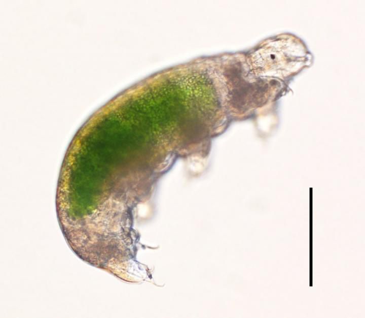
| 22:21, 8 December 2022 | 109006 web.jpg (file) |  |
26 KB | Spivack2 | cute transparent eutardigrade light microscope | 1 |
| 14:19, 9 June 2006 | 10x.jpg (file) |  |
68 KB | WikiAdmin | 1 | |
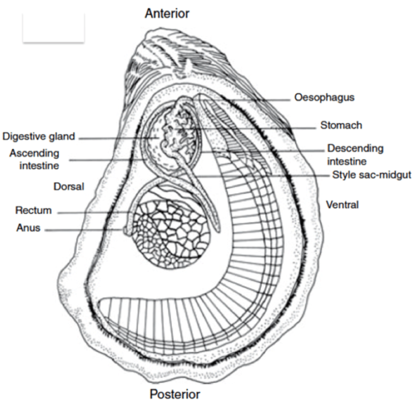
| 13:27, 8 November 2019 | 11-The-digestive-system-of-Eastern-oyster-C-virginica-Picture-is-adapted-from-Gosling.png (file) |  |
229 KB | Hamilton5 | 1 | |
| 04:15, 4 December 2015 | 111.jpeg (file) |  |
251 KB | Kaleen.timon | 1 | |
| 17:46, 3 May 2012 | 111111.jpg (file) |  |
8 KB | Mujyanamaf | 1 | |
| 04:18, 4 December 2015 | 112.jpeg (file) |  |
426 KB | Kaleen.timon | 1 | |
| 04:24, 4 December 2015 | 113.jpeg (file) |  |
495 KB | Kaleen.timon | 1 | |
| 04:25, 4 December 2015 | 114.jpeg (file) |  |
615 KB | Kaleen.timon | 1 | |
| 04:29, 4 December 2015 | 115.jpeg (file) |  |
230 KB | Kaleen.timon | 1 | |
| 04:34, 4 December 2015 | 116.jpeg (file) |  |
373 KB | Kaleen.timon | 1 | |
| 04:37, 4 December 2015 | 117.jpeg (file) |  |
423 KB | Kaleen.timon | 1 | |
| 12:59, 4 December 2015 | 118.jpeg (file) |  |
46 KB | Kaleen.timon | 1 | |
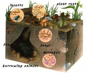
| 22:42, 14 March 2016 | 11 living soil.jpg (file) |  |
18 KB | Kjfyhrie | 1 | |
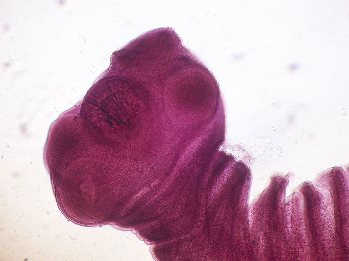
| 15:28, 5 April 2017 | 1200px-Taenia solium scolex.jpeg (file) |  |
122 KB | Headj | Scolex (head) of Taenia solium | 1 |
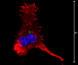
| 16:02, 7 November 2013 | 120126-dendritic-cell-bcg-iah-300.jpg (file) |  |
47 KB | Treimans | 1 | |
| 15:22, 11 April 2024 | 123.png (file) |  |
137 KB | Abramson1 | 1 | |
| 00:27, 15 April 2024 | 123123123.jpeg (file) |  |
39 KB | Li13 | 1 | |
| 19:48, 2 December 2016 | 125376.jpg (file) |  |
65 KB | Bmjones6353 | 1 | |
| 20:15, 7 October 2011 | 12563 lores.jpg (file) |  |
29 KB | Ziegler.s | A photomicrograph of mature Plasmodium malariae schizont in an infected RBC. Content Providers: CDC/Dr. Mae Melvin This media comes from the Centers for Disease Control and Prevention's Public Health Image Library (PHIL), with identification number #27 | 1 |
| 03:59, 4 March 2016 | 125980780.jpg (file) |  |
11 KB | Dkeat | 1 | |
| 22:24, 6 December 2019 | 1280 0x6eFJ8729pL.jpg (file) |  |
297 KB | Hamilton5 | 1 | |
| 03:24, 7 December 2019 | 12864 2014 6923 Fig5 HTML.jpg (file) |  |
57 KB | Jones6 | 1 | |
| 16:06, 18 November 2011 | 1297-9716-42-102-1.jpg (file) |  |
117 KB | Maggiebenson | 1 | |
| 09:07, 28 May 2007 | 1298219234.jpg (file) |  |
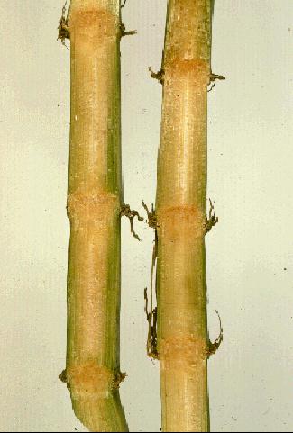
24 KB | Csl002 | Discoloration of sugarcane vascular bundles caused by ratoon stunting disease. | 1 |
