File list
From MicrobeWiki, the student-edited microbiology resource
This special page shows all uploaded files.
| Date | Name | Thumbnail | Size | User | Description | Versions |
|---|---|---|---|---|---|---|
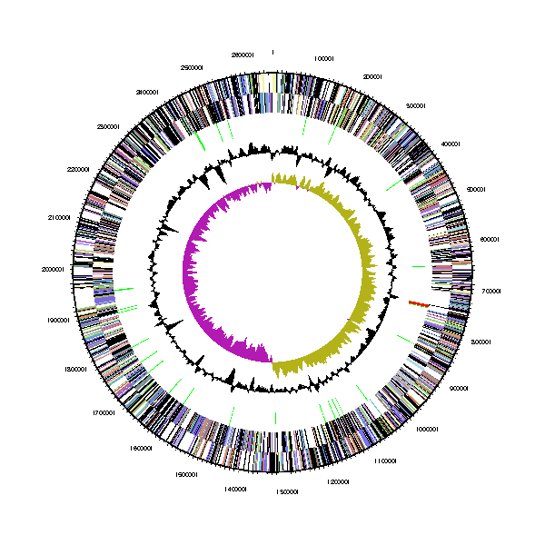
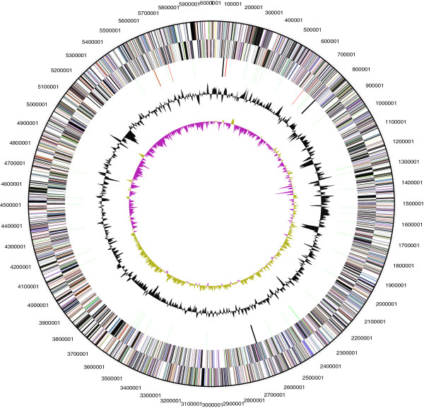
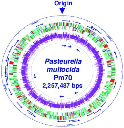
| 07:26, 2 April 2017 | Bdellovibrio exovorus chromosome map.png (file) |  |
40 KB | PierceK | Chromosome map of circular chromosome of bdellovibrio exovorus strain JSS. From outside to the center: Genes on forward strand (color by COG categories) Genes on reverse strand (color by COG categories) RNA genes (tRNAs green, rRNAs red, other RNAs bl... | 1 |
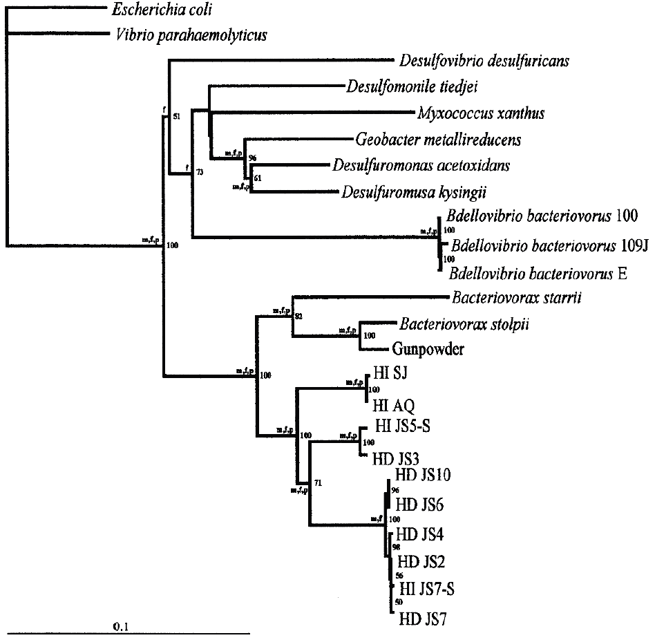
| 07:12, 2 April 2017 | Bd bacteriovorus phylogenetic tree.png (file) |  |
85 KB | PierceK | A neighbor-joining tree based on 16SrRNA sequences demonstrating the phylogenetic relationship between marine and terrestrial Bdellovibrio isolates. Taken from "Bergey's Manual of Systematic Bacteriology" Vol. 2 part C (2nd Ed.), p.1050 | 1 |
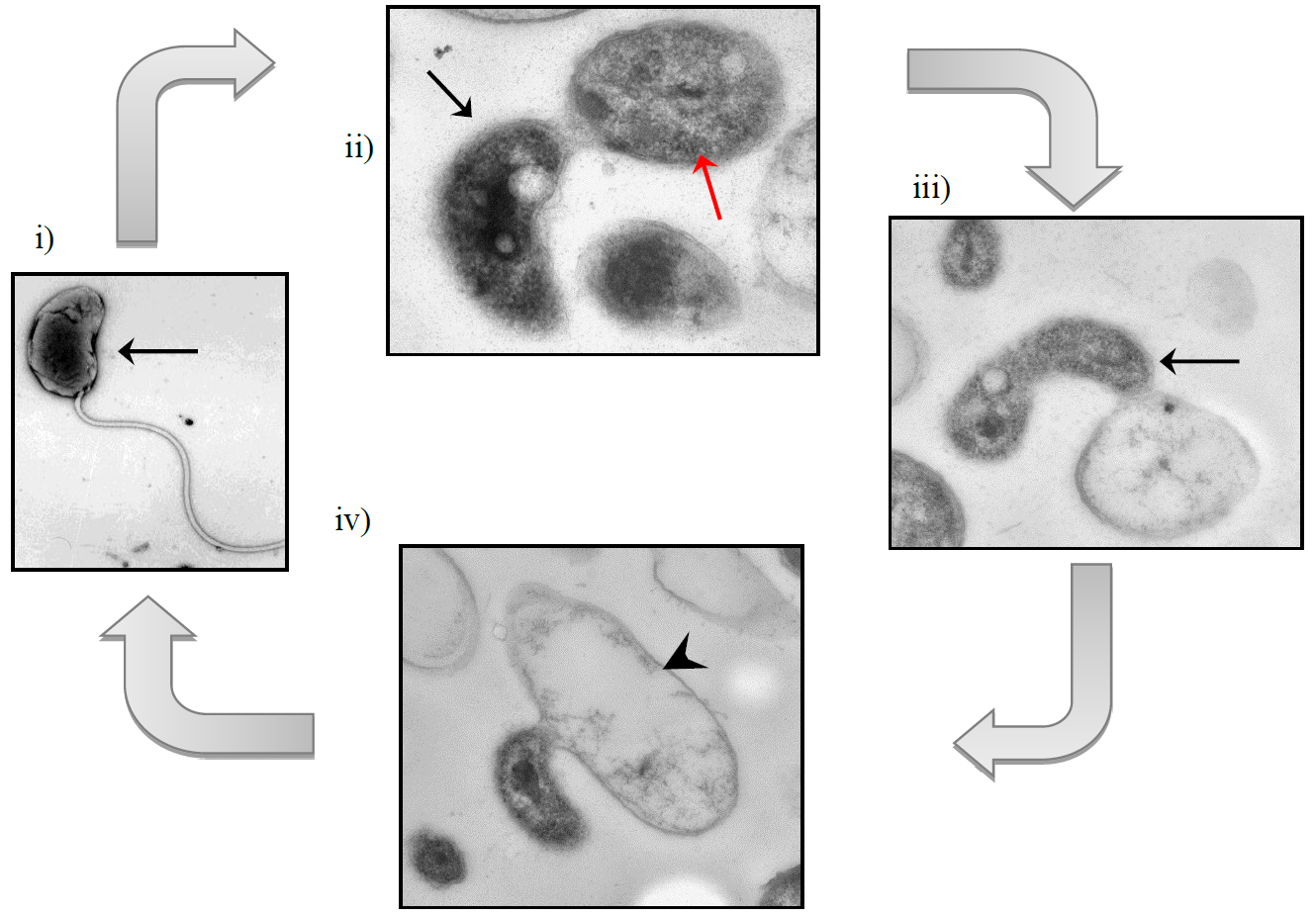
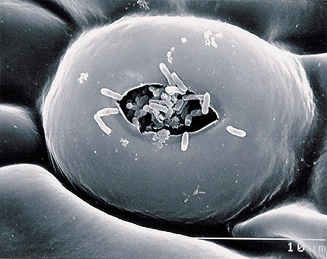
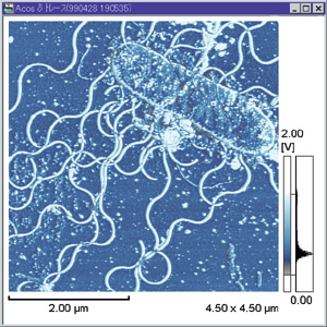
| 06:32, 2 April 2017 | Bdellovibrio exovorus epibiotic life cycle.png (file) |  |
336 KB | PierceK | Epibiotic life cycle of Bd. exovorus. i) Motile attack phase cell (black arrow); ii) Attachment of Bd. exovorus to prey cell, Stenotrophomonas maltophilia (red arrow); iii) Replication of Bd. exovorus by binary fission; iv) Prey cell cytoplasmic conten... | 1 |
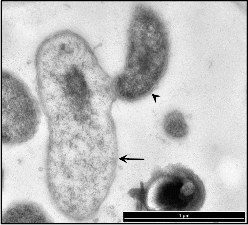
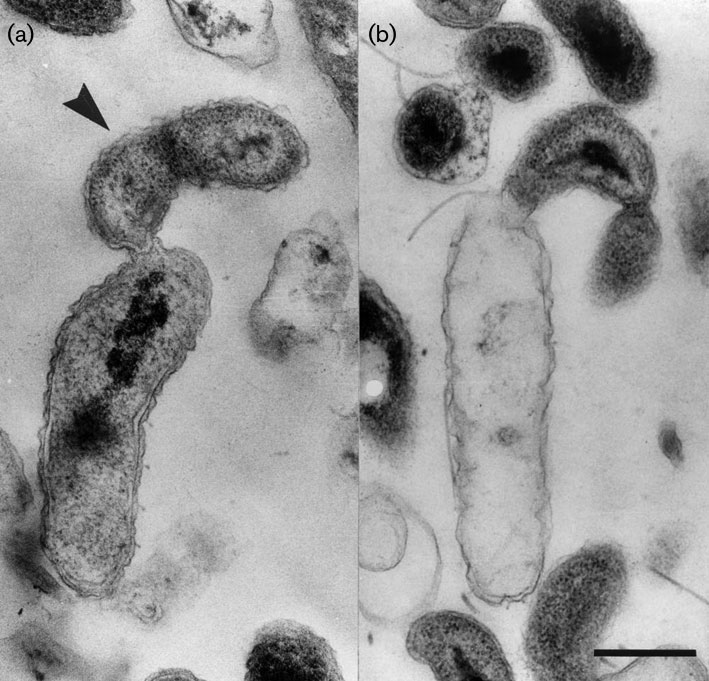
| 06:22, 2 April 2017 | Bdellovibrio exovorus on Stenotrophomonas maltophilia.png (file) |  |
636 KB | PierceK | Epibiotic life cycle of Bd. exovorus FFRS-5 confirmed by thin section transmission electron microscopy. Thin section of Bd. exovorus FFRS-5 (black arrowhead) at the surface of S. maltophilia (black arrow)cell during predation. A small amount of cytopl... | 1 |
| 20:35, 31 March 2017 | Vibalgdis.jpg (file) |  |
59 KB | MorrisP | 3 | |
| 19:02, 31 March 2017 | Vibalgpol.jpg (file) |  |
31 KB | MorrisP | 2 | |
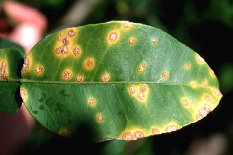
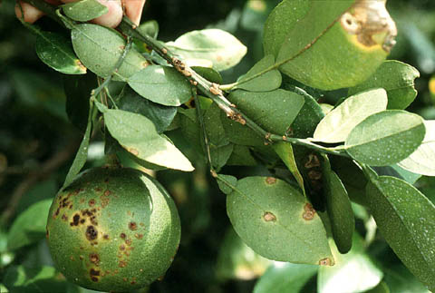
| 05:46, 30 March 2017 | Xcitri.jpg (file) |  |
136 KB | JangY | 1 | |
| 05:27, 30 March 2017 | CitrusCanker03.jpg (file) |  |
31 KB | JangY | 1 | |
| 05:26, 30 March 2017 | CitrusCanker01.jpg (file) |  |
41 KB | JangY | 1 | |
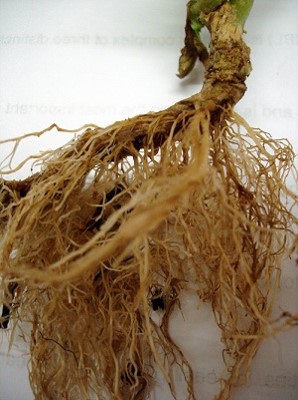
| 03:00, 30 March 2017 | Imagen1.jpg (file) |  |
39 KB | TamezI | Tobacco Plant Showing Symptoms of Hairy Root Disease Photo Credit: Adriana M. Allippi, Facultad de Ciencias Agrarias y Forestales, Argentina. | 1 |
| 01:10, 30 March 2017 | Brasiliensis.jpeg (file) |  |
6 KB | GallegosF | 1 | |
| 00:57, 30 March 2017 | 40793 2014 Article 10 Fig3 HTML.jpg (file) |  |
124 KB | GallegosF | 1 | |
| 00:55, 30 March 2017 | Genome and Sequencing.jpeg (file) |  |
124 KB | GallegosF | 1 | |
| 23:12, 29 March 2017 | Dsc 0483.jpg (file) |  |
103 KB | TamezI | Agrobacterium Rhizogenes, Gram stain 1000X magnification, Brightfield Microscopy | 1 |
| 12:05, 22 March 2017 | Pasturella multicida.jpeg (file) |  |
40 KB | PlaceJ | 1 | |
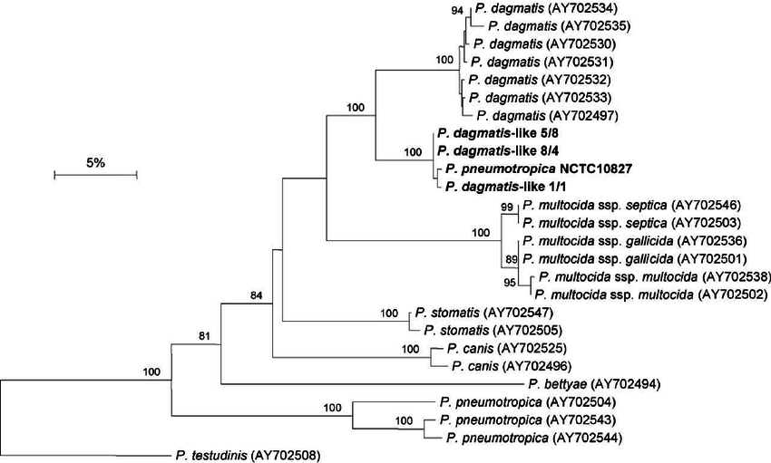
| 11:47, 22 March 2017 | Pasturella phylogenetic tree.png (file) |  |
69 KB | PlaceJ | 2 | |
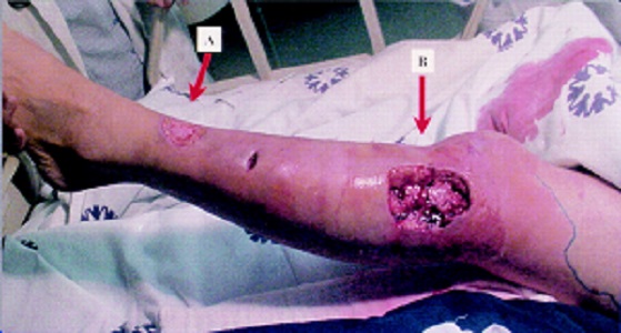
| 17:30, 25 February 2017 | Pasteurella-canis.JPG (file) |  |
89 KB | PlaceJ | 1 | |
| 21:05, 19 February 2017 | Pasteurella-multocida.JPG (file) |  |
216 KB | PlaceJ | 1 | |
| 04:14, 14 February 2017 | Vibalg.jpg (file) |  |
54 KB | MorrisP | 1 | |
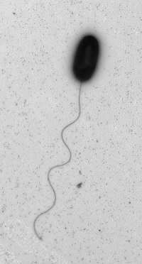
| 09:01, 13 February 2017 | Bdellovibrio exovorus.png (file) |  |
288 KB | PierceK | Fig. 1. Thin sections of cells of strain JSST attached to intact (a) and empty (b) stalked cells of Caulobacter crescentus CB2A. The arrowhead in (a) indicates the cell of strain JSST. Bar, 500 nm. The image in (a) is reprinted from the cover of Journa... | 1 |
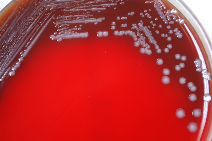
| 17:59, 11 February 2017 | Pasturella Culture.JPG (file) |  |
33 KB | PlaceJ | 1 | |
| 02:57, 13 December 2016 | Odoribacter Electron Micrograph.jpg (file) |  |
106 KB | Cecthai | 1 | |
| 23:39, 12 December 2016 | Odoribacter Electron Micrograph.gif (file) |  |
657 KB | Cecthai | 1 | |
| 21:43, 12 December 2016 | Tempeh.jpg (file) |  |
41 KB | Hkim1007 | 1 | |
| 18:06, 12 December 2016 | Figure 3.jpeg (file) |  |
10 KB | Jbonet13 | 3 | |
| 18:04, 12 December 2016 | Figure 2.jpeg (file) |  |
35 KB | Jbonet13 | 3 | |
| 17:56, 12 December 2016 | Figure 1.png (file) |  |
316 KB | Jbonet13 | Animated visual of genus Eubacterium cell structure. | 3 |
| 17:54, 12 December 2016 | Figure 1.pdf (file) | 102 KB | Jbonet13 | Animated visual of genus Eubacterium cell structure. | 1 | |
| 15:39, 12 December 2016 | Leucobacter Phylogeny 2.png (file) |  |
32 KB | Cvt | 2 | |
| 15:25, 12 December 2016 | P.palmivora 3.jpg (file) |  |
9 KB | Garciay | 1 | |
| 15:25, 12 December 2016 | P.palmivora 2.jpg (file) |  |
10 KB | Garciay | 1 | |
| 15:17, 12 December 2016 | P.palmivora 1.jpg (file) |  |
12 KB | Garciay | 1 | |
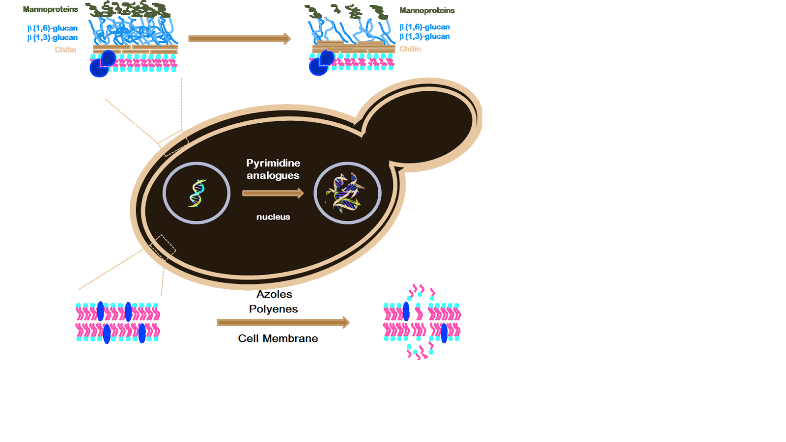
| 15:14, 12 December 2016 | Mechanism of Antifungal Resistance.png (file) |  |
217 KB | Kdoiron | 1 | |
| 15:09, 12 December 2016 | C krusei.png (file) |  |
1.8 MB | Kdoiron | 1 | |
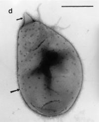
| 15:06, 12 December 2016 | Nematopsis2.gif (file) |  |
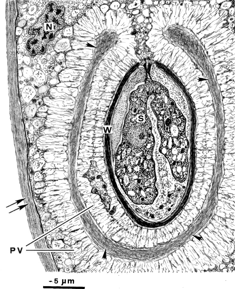
764 KB | Suppleej | Schematic drawing of a longitudinal section of an oocyst of Nematopsis gigas n. sp. and the surrounding structures of the parasito-phorous vacuole (PV). Note the numerous anastomosing microfibrils of the PV, some of which form a dense and complex netwo... | 1 |
| 15:05, 12 December 2016 | Nematopsis1.gif (file) |  |
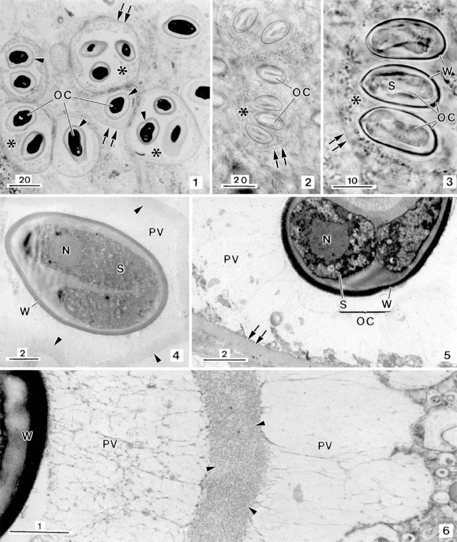
876 KB | Suppleej | Light microscopy of the apicomplexan gregarine, Nematopsis gigas n. sp., a parasite found in the mantle tissue of the gastropod Nerita ascencionis (all scale bars in μm). Semithin section showing some host phagocytes (*) each containing some oocysts (... | 2 |
| 14:51, 12 December 2016 | Figure 1 - Genome Structure.png (file) |  |
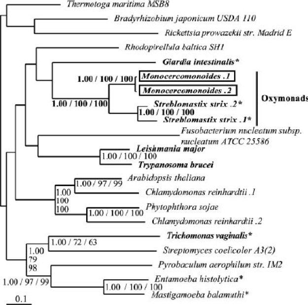
200 KB | Slcarpen | Phylogenetic tree of excavates and closely related amoeba. Excavates include <i>Monocercomonoides</i>, <i>Giardia</i>, and <i>Trichomonas</i>. By construction with pyruvate phosphate dikinase (PPDK), it is evident that <i>Monocercomonoides</i> is most ... | 1 |
| 14:47, 12 December 2016 | Worm Star.png (file) |  |
73 KB | Cvt | 1 | |
| 14:45, 12 December 2016 | Figure 4 - Metabolism.png (file) |  |
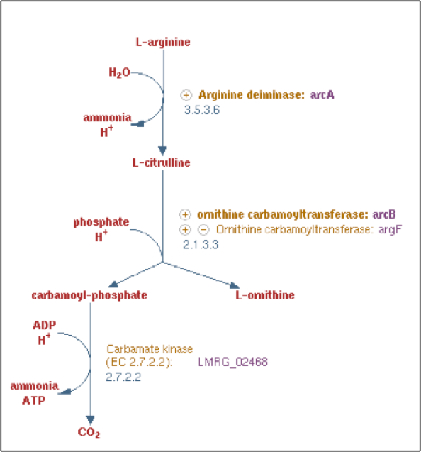
75 KB | Slcarpen | Arginine degradation/deiminase pathway similar to that present in <i>Monocercomonoides</i>. Important enzymes include arginine deiminase, ornithine carbomoyltransferase, and carbamate kinase. Important products include ATP, inorganic phosphate, and amm... | 1 |
| 14:44, 12 December 2016 | Leucobacter Phylogeny 1.png (file) |  |
56 KB | Cvt | 1 | |
| 14:43, 12 December 2016 | Figure 3 - Metabolism.png (file) |  |
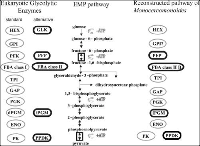
203 KB | Slcarpen | Comparison of <i>Monocercomonoides</i> glycolytic pathway with that of a typical eukaryote. Enzymes outlined in bold represent changes from the typical pathway. Reprinted with permission from Liapounova et al., 2006. | 1 |
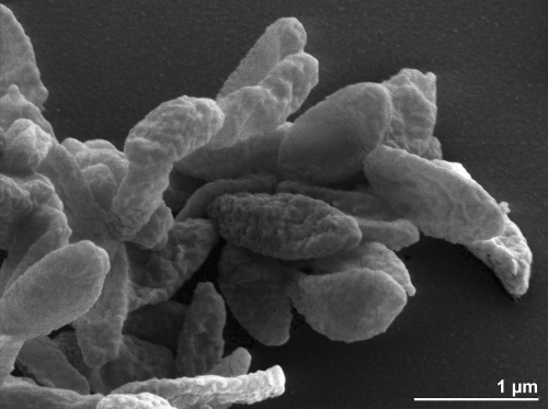
| 14:43, 12 December 2016 | Cryptococcus gattii 02.jpg (file) |  |
53 KB | Armal629 | 1 | |
| 14:38, 12 December 2016 | Cell Light Micrograph Figure 2.png (file) |  |
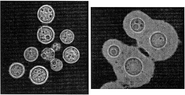
65 KB | Slcarpen | Light Micrograph of <i>Monocercomonoides</i>. The light micrograph images depict the grouping of flagella, which are all longer than the body, on each side of an anterior nucleus. | 1 |
| 05:11, 9 December 2016 | IndexGraphic226.jpg (file) |  |
27 KB | Thomas.dominguez1 | 1 | |
| 05:09, 9 December 2016 | Micrococcus 2.jpg (file) |  |
3 KB | Thomas.dominguez1 | 1 | |
| 04:26, 9 December 2016 | Saltbc.jpg (file) |  |
1.79 MB | Carley.cudmore | 1 | |
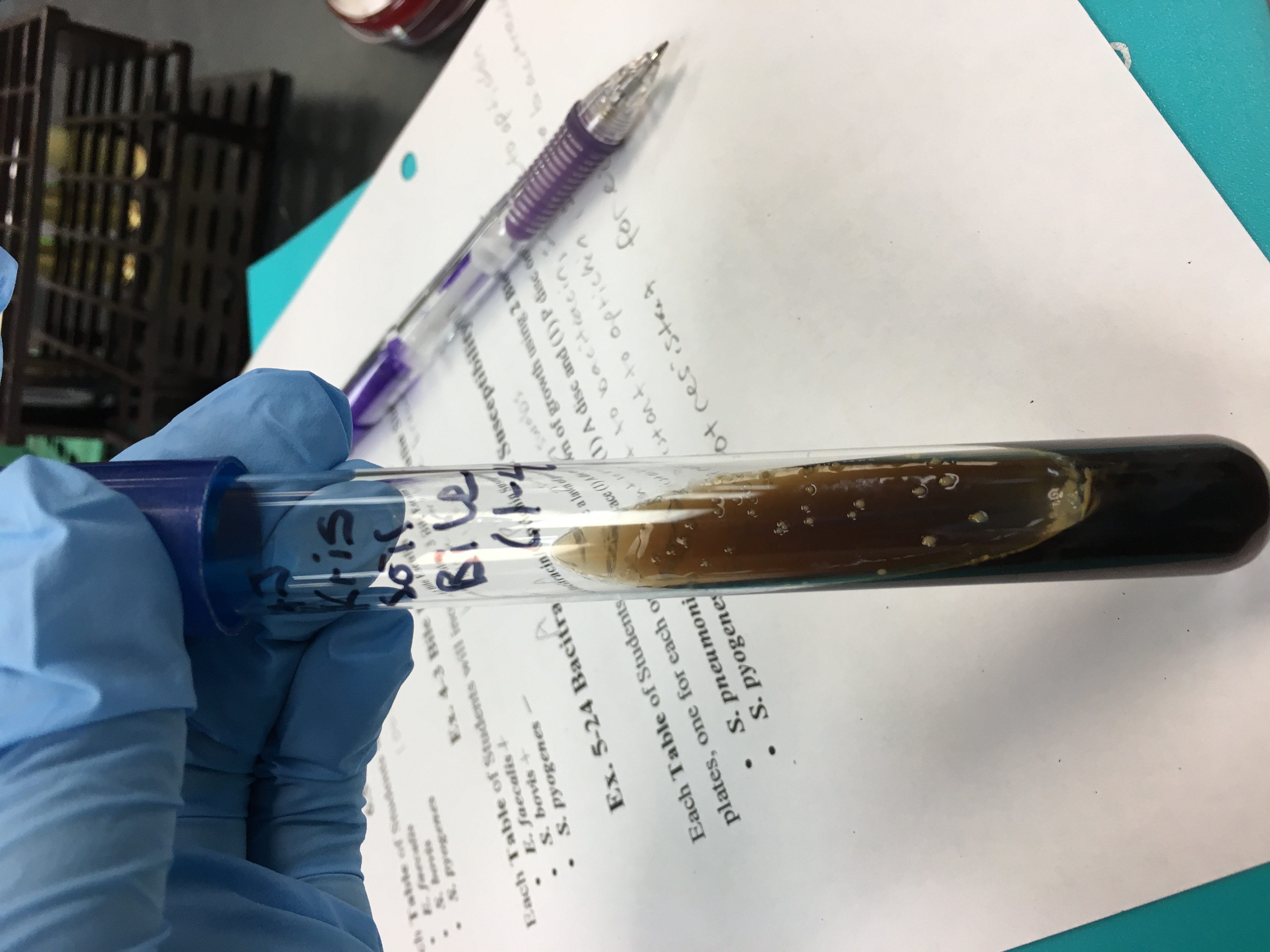
| 04:20, 9 December 2016 | Bile.jpg (file) |  |
1.61 MB | Carley.cudmore | 1 | |
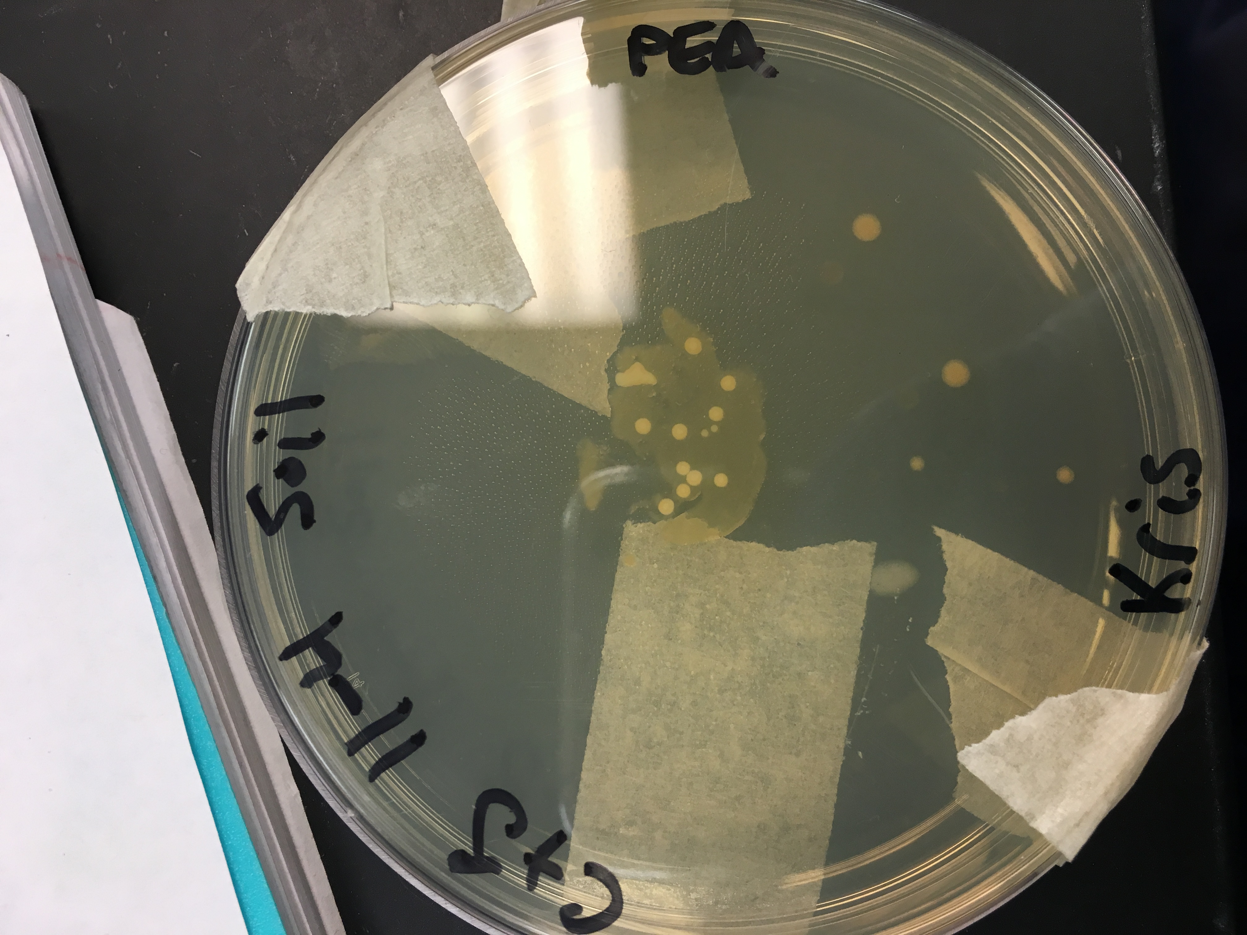
| 04:16, 9 December 2016 | Pea.jpg (file) |  |
1.99 MB | Carley.cudmore | 1 | |
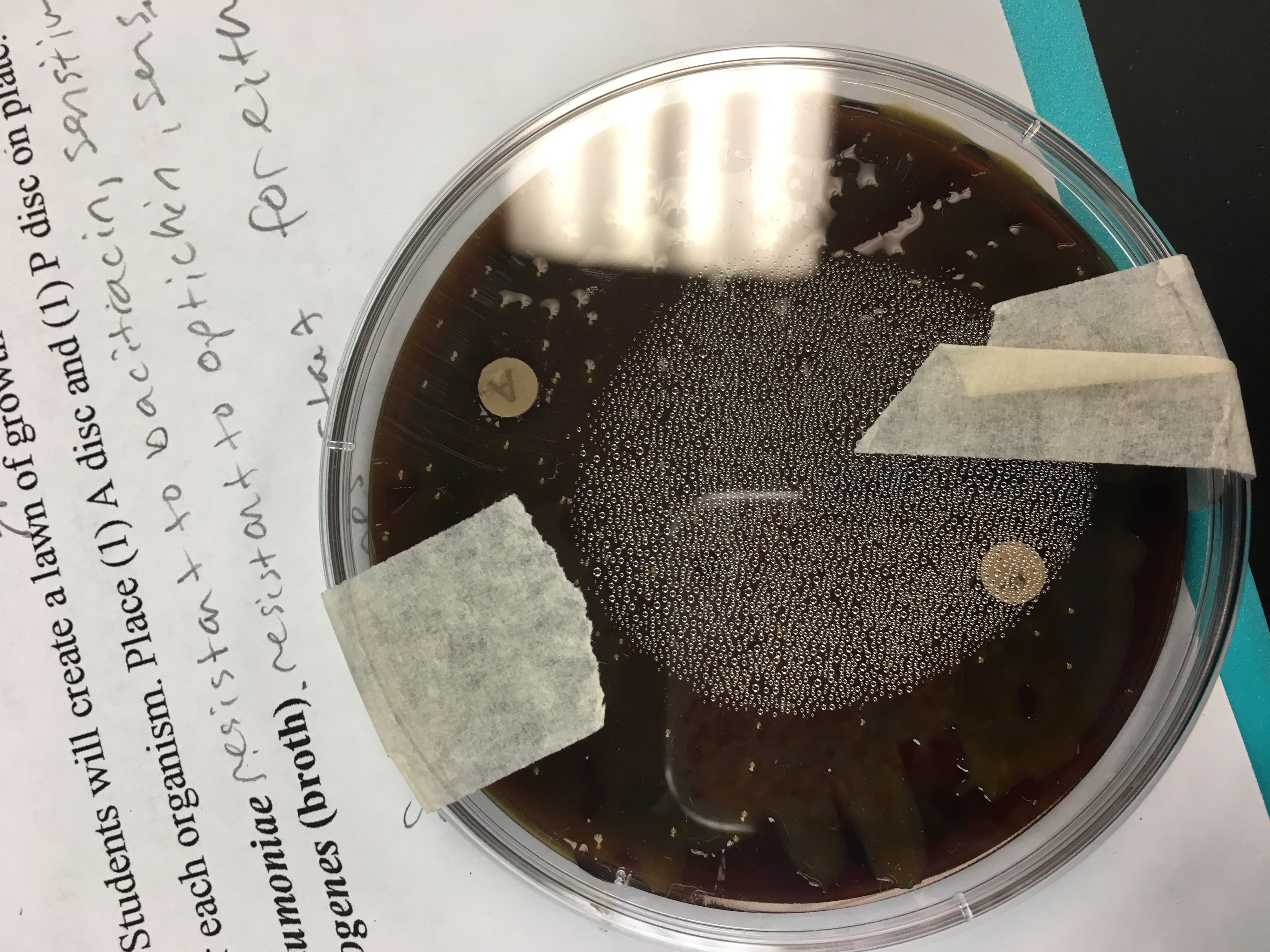
| 04:12, 9 December 2016 | Ap2.jpg (file) |  |
2.57 MB | Carley.cudmore | 1 | |
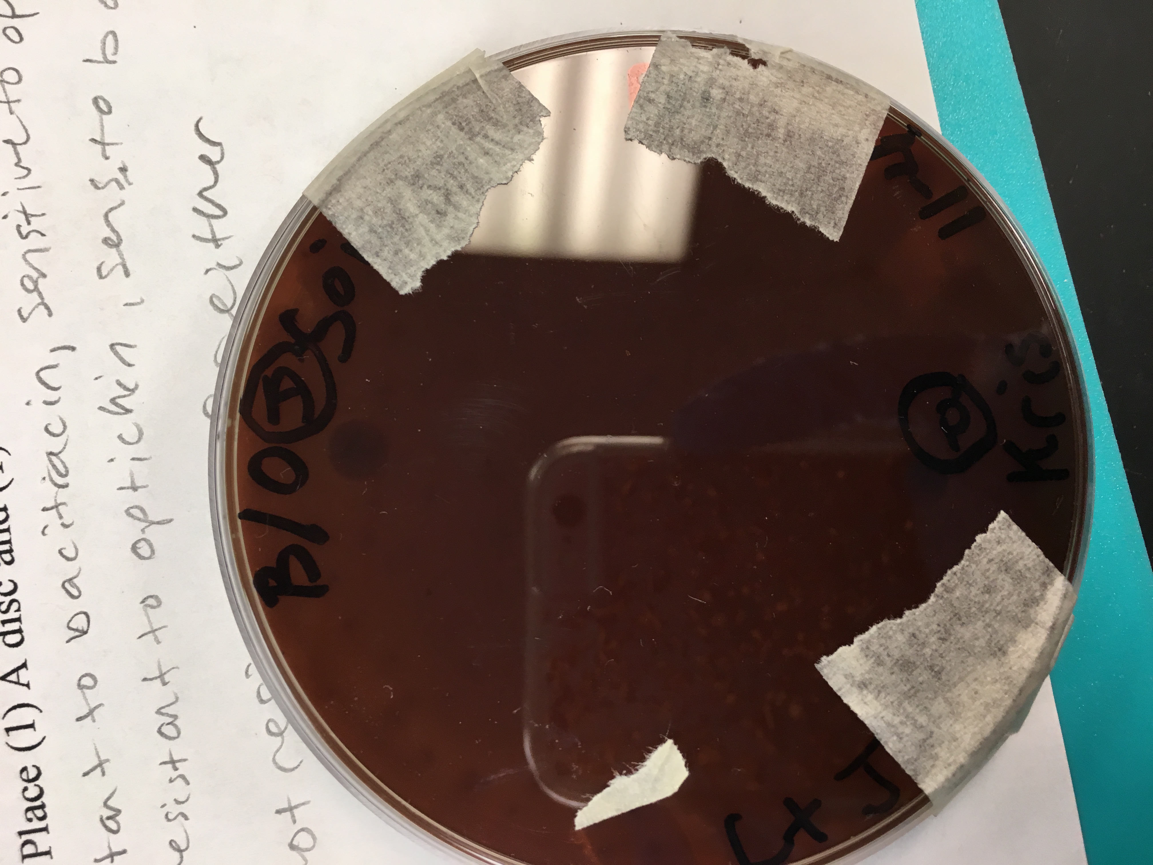
| 04:11, 9 December 2016 | Ap1.jpg (file) |  |
1.88 MB | Carley.cudmore | 1 |
